
Page 12 - Read Online
P. 12
Bansberg et al. Plast Aesthet Res 2024;11:12 https://dx.doi.org/10.20517/2347-9264.2023.109 Page 7 of 13
Despite substantial experience in closing perforations, attempted repair remains challenging when prior
septal surgery has been performed regardless of the perforation size. Septal scarring due to one or more
prior procedures can substantially impact the ability to develop surgical planes for intact mucosal elevation.
The removal of bone and cartilage during septal surgery results in densely adherent and attenuated mucosa
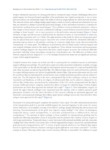
that can extend for a distance beyond the perforation margin. A slow and tedious dissection is common for
perforations resulting from prior septal surgery. This condition is most problematic posteriorly, where a
thin margin of fused mucosa can pose a substantial challenge to intact bilateral separation before reaching
cartilage or bone located 1 cm or more posterior to the perforation mucosal margin [Figure 4]. Intact
elevation of right and left mucosa is facilitated by the injection of saline or local anesthetic to widen the
margin sharp separation with a #15 blade. The eight patients in this study for whom an interposition graft
was not placed had prior septal surgery. Operative note review described the substantial difficulty in
elevating mucosa intact in all patients, resulting in a tenuous single-layer repair that prevented the
“interposing” of a tissue graft. The difference in perforation sizes for failed repairs between surgical and
non-surgical etiologies noted in this study was significant. These clinical observations and measurement
analysis findings support our impression that prior septal surgery increases the technical difficulty
associated with flap closure procedures, irrespective of perforation size. The difference in failure rates
between surgical and non-surgical (5.7 vs. 4.7%) etiologies found in this study was not significant and may,
in part, reflect surgical experience.
Complete tension-free closure on at least one side is a prerequisite for consistent success in perforation
surgery utilizing mucosal flaps. The perforation repair procedure presented emphasizes complete coverage
of the septal defect on the left side through the development and advancement of a superior bipedicled flap
and, when necessary, an inferior bipedicled flap. The elliptical shape noted for most perforations conforms
to a repair utilizing superior and inferior longitudinally oriented flaps. Flap incision placement and the need
for an inferior flap are determined by several factors, most notably perforation position and size relative to
nasal size. The left superior flap is the most consequential flap in this technique owing to its robust
vascularity and thickness, as well as the degree of advancement that can be achieved compared to the
inferior flap. Furthermore, mucosa from the undersurface of the ULC can be incorporated into the flap to
increase width and achieve complete, tension-free defect coverage for relatively larger (vertical height)
[16]
perforations and those that approach the internal valve angle [Figure 5]. Prior rhinoplastic surgery, in
which the upper lateral cartilages were separated from the septum, with or without spreader graft
placement, interferes with the intact elevation of ULC mucosa utilizing an endonasal approach. An
alternative, non-incisional technique that utilizes upper lateral cartilage mucosa has been described when
[5]
using an open approach for the repair [Figure 6].
Placement of an interposition graft completes the standard 3-layer repair. The often-stated primary function
of the interposition graft is to provide scaffold support for mucosal migration in the event of a suture
closure dehiscence or incomplete coverage of the defect’s cartilage margin following flap advancement and
suturing. Neovascularization of the graft may improve repair healing . Our choice of autologous
[12]
temporalis fascia, septal cartilage/bone, or auricular perichondrium is influenced by clinical
circumstances . The amount of septal cartilage or bone available for interpositional grafting may be
[17]
insufficient following septoplasty. Fascia is frequently used for patients with a history of prior surgery or
with larger perforations. Temporalis fascia is easily harvested with minimal morbidity and risk. Auricular
cartilage that is harvested for concurrent valve or dorsal augmentation surgery can provide a perichondrium
[18]
graft and avoid a temporal donor site . A review of graft material selection in our bilateral flap repairs
[17]
found no significant difference between autologous graft application and closure outcomes . Another
attempt at closure following a failed repair can be extremely difficult, or impossible. The addition of a graft