
Page 13 - Read Online
P. 13
Page 8 of 13 Bansberg et al. Plast Aesthet Res 2024;11:12 https://dx.doi.org/10.20517/2347-9264.2023.109
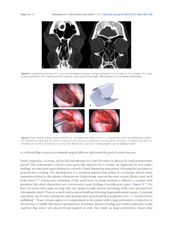
Figure 4. Computed tomography (CT) scan coronal images of surgical etiology perforation 12 mm length by 9 mm height. (A) Image
through perforation; (B) Image through thin posterior margin absent of cartilage; Patient opted for a customized septal button.
Figure 5. Upper lateral cartilage mucosa (ULM) flap. (A) Endoscopic photo of 1.5 × 1.2 cm perforation and intercartilaginous incision;
(B) Dashed lines depicting the extent of elevation and incisions connecting to dorsal septum incisions; (C) Endoscopic photo of
elevation; (D) ULM flap advanced for suturing; Note the thin ULC mucosa (*) overlapping the superior cartilage margin.
to a bilateral flap repair poses minimal surgical effort or risk toward the goal of closure success.
Nasal congestion, crusting, and palatal paresthesias are noted by patients during the early postoperative
period. The postoperative clinical course generally stabilizes by 6 months. As supported by this study’s
findings, we note most repair failures by 6 months. Nasal obstruction may persist following the resolution of
postoperative swelling. The development of a unilateral superior flap results in a technique-driven repair
asymmetry related to the inferior advancement of physiologic mucosa that may contain thicker nasal swell
body tissue [19,20] . Submucosal reduction of the swell body by sharp excision is offered to patients with
persistent left-sided obstruction and corroborative exam findings 6 months post repair [Figure 7] . We
[19]
have not noted valve angle scarring with our repairs, though inferior narrowing of the valve area has been
infrequently noted. There is a small risk for dorsal saddling following larger perforation repairs. A supratip
depression can be noted during the early postoperative period and then progresses over 3-4 months before
stabilizing . Nasal dorsum support is compromised in the patient with a large perforation, irrespective of
[16]
the presence of saddle deformity at presentation. Secondary intention healing and wound contracture at the
superior flap donor site places dorsal support at risk. Our study on large perforation repairs that