
Page 63 - Read Online
P. 63
Patel et al. Plast Aesthet Res 2024;11:20 https://dx.doi.org/10.20517/2347-9264.2024.17 Page 9 of 14
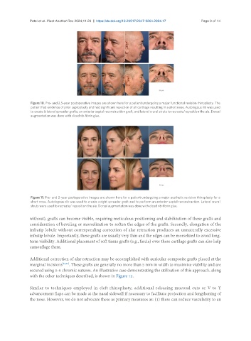
Figure 10. Pre- and 2.5-year postoperative images are shown here for a patient undergoing a major functional revision rhinoplasty. The
patient had evidence of prior septoplasty and had significant resection of all cartilage resulting in a short nose. Autologous rib was used
to create bilateral spreader grafts, an anterior septal reconstruction graft, and lateral crural struts to recreate/reposition the ala. Dorsal
augmentation was done with diced rib-fibrin glue.
Figure 11. Pre- and 2-year postoperative images are shown here for a patient undergoing a major aesthetic revision rhinoplasty for a
short nose. Autologous rib was used to create a right spreader graft and to perform an anterior septal reconstruction. Lateral crural
struts were used to recreate/ reposition the ala. Dorsal augmentation was done with diced rib-fibrin glue.
without), grafts can become visible, requiring meticulous positioning and stabilization of these grafts and
consideration of beveling or morselization to soften the edges of the grafts. Secondly, elongation of the
infratip lobule without corresponding correction of alar retraction produces an unnaturally excessive
infratip lobule. Importantly, these grafts are usually very thin and the edges can be morselized to avoid long-
term visibility. Additional placement of soft tissue grafts (e.g., fascia) over these cartilage grafts can also help
camouflage them.
Additional correction of alar retraction may be accomplished with auricular composite grafts placed at the
marginal incisions [32,33] . These grafts are generally no more than 5 mm in width to maximize viability and are
secured using 5-0 chromic sutures. An illustrative case demonstrating the utilization of this approach, along
with the other techniques described, is shown in Figure 12.
Similar to techniques employed in cleft rhinoplasty, additional releasing mucosal cuts or V to Y
advancement flaps can be made at the nasal sidewall if necessary to facilitate projection and lengthening of
the nose. However, we do not advocate these as primary measures as: (1) these can reduce vascularity to an