
Page 59 - Read Online
P. 59
Patel et al. Plast Aesthet Res 2024;11:20 https://dx.doi.org/10.20517/2347-9264.2024.17 Page 5 of 14
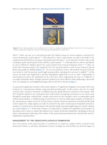
Figure 3. Costal cartilage grafts should be obliquely cut to minimize warping and subsequently left in saline for 30 min to assess for
inherent warping. This is especially important when using long cartilage grafts, as shown here.
[15]
SEGs . Grafts can run in an extended spreader-like fashion along the dorsal septum or primarily be
anchored along the caudal septum [13,15] . SEGs placed in a side-to-side manner can add to the width of the
caudal septum, but this does not impact functional nasal outcomes . As an alternative to these side-to-side
[16]
techniques, grafts may be placed in line with the caudal septum [17,18] . Grafts placed in an end-to-end fashion
are more difficult to stabilize against the caudal septum with suturing techniques alone [15,18,19] . Thus, we
prefer such extension grafts to be supported with two spreader grafts as well as more inferiorly placed
[20]
batten-type grafts for stability [Figure 4] . In a recent systematic review, this method of septal extension
was shown to yield the greatest nasal length among studies reporting this outcome (8 studies) . In this
[21]
review, the mean nasal length before and after rhinoplasty ranged from 0.28 to 6.2 mm . Importantly, as
[21]
aforementioned, given the limitations of the soft tissue when lengthening the nose, it is difficult to
accurately determine which cartilage extension method is the most effective when addressing a shortened
nose. An example of a nose lengthened using a side-to-side SEG is shown in Figure 5.
If a patient has significant retrusion of the native septum or if significant elongation is necessary, SEGs may
be placed in a discontinuous fashion using extended spreader grafts. In this scenario, the use of a septal
extension that is fixated to bilateral or unilateral spreader grafts allows for extension of the septum. This
SEG should be fixated to the nasal spine either with sutures through the periosteum or via a hole made
through the nasal spine. Alternatively, a wedge can be made into the maxillary crest to accommodate a graft
without suture fixation. A similar strategy is used if there is a loss of caudal septal integrity or deviations of
the caudal septum require resection. In this scenario, anterior septal reconstruction is needed and the graft
used to replace the caudal septum can either be sutured to the native dorsal septum if minimal extension is
needed or be fixated to the dorsal septum with spreader grafts if greater lengthening and counter rotation
are needed [Figure 6] . In the latter scenario, it is important to consider the angulation and projection of
[22]
the grafts used to replace or extend the septum in the context of soft tissue limitations. Redraping of the soft
tissue and temporary anchoring of the domes to the newly positioned septum may elucidate the need for
adjustments to graft position.
MANAGEMENT OF THE OSSEOCARTILAGINOUS FRAMEWORK
Once the position of the septal structure is established, the nasal tip complex will be attached to this
framework. Similar to soft tissue, limitations in lateral cartilage size and mobility can limit the degree of