
Page 65 - Read Online
P. 65
Patel et al. Plast Aesthet Res 2024;11:20 https://dx.doi.org/10.20517/2347-9264.2024.17 Page 11 of 14
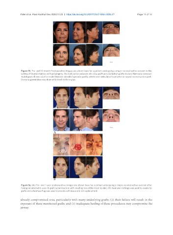
Figure 15. Pre- and 10-month Postoperative images are shown here for a patient undergoing a major reconstructive revision in the
setting of Granulomatosis with polyangiitis. She had a prior cadaveric rib onlay graft and alar batten grafts in place that were removed.
Autologous rib was used to create bilateral extended spreader grafts, which were articulated to an anterior septal reconstruction graft.
Dorsal augmentation was done with diced rib-fibrin glue.
Figure 16. (A) Pre- and 1-year postoperative images are shown here for a patient undergoing a major reconstructive revision after
having an infected K-wire rib graft reconstruction with resulting loss of his nasal tip skin; (B) Auricular cartilage was used to create tip
grafts and a forehead flap was used to provide soft tissue and skin replacement.
already compromised area, particularly with many underlying grafts; (2) their failure will result in the
exposure of these mentioned grafts; and (3) inadequate healing of these procedures may compromise the
airway.