
Page 19 - Read Online
P. 19
Gunderson et al. Plast Aesthet Res 2023;10:50 https://dx.doi.org/10.20517/2347-9264.2023.42 Page 13 of 15
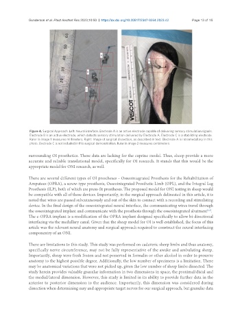
Figure 4. Surgical Approach. Left: Neural interface. Electrode A is an active electrode capable of delivering sensory stimulation signals.
Electrode B is an active electrode, which detects sensory stimulation delivered by Electrode A. Electrode C is a stabilizing electrode.
Ruler in image 1 measures millimeters. Right: Image of surgical dissection, as described in text. Electrode A is intramedullary in this
photo. Electrode C is not included in this surgical demonstration. Ruler in image 2 measures centimeters.
surrounding OI prosthetics. These data are lacking for the caprine model. Thus, sheep provide a more
accurate and reliable translational model, specifically for OI research. It stands that this would be the
appropriate model for ONI research, as well.
There are several different types of OI prostheses – Osseointegrated Prosthesis for the Rehabilitation of
Amputees (OPRA), a screw-type prosthesis, Osseointegrated Prosthetic Limb (OPL), and the Integral Leg
Prosthesis (ILP), both of which are press-fit prostheses. The proposed model for ONI testing in sheep would
be compatible with all of these devices. Importantly, in the surgical approach delineated in this article, it is
noted that wires are passed subcutaneously and out of the skin to connect with a recording and stimulating
device. In the final design of the osseointegrated neural interface, the communicating wires travel through
the osseointegrated implant and communicate with the prosthesis through the osseointegrated abutment .
[12]
The e-OPRA implant is a modification of the OPRA implant designed specifically to allow bi-directional
interfacing via the medullary canal. Given that the sheep model for OI is well established, the focus of this
article was the relevant neural anatomy and surgical approach required to construct the neural interfacing
componentry of an ONI.
There are limitations to this study. This study was performed on cadaveric sheep limbs and thus anatomy,
specifically nerve circumference, may not be fully representative of the awake and ambulating sheep.
Importantly, sheep were fresh frozen and not preserved in formalin or other alcohol in order to preserve
anatomy to the highest possible degree. Additionally, the low number of specimens is a limitation. There
may be anatomical variations that were not picked up, given the low number of sheep limbs dissected. The
study herein provides valuable granular information in two dimensions in space, the proximal/distal and
the medial/lateral dimension. However, this study is limited in its ability to provide further data in the
anterior to posterior dimension to the audience. Importantly, this dimension was considered during
dissection when determining easy and appropriate target nerves for our surgical approach, but granular data