
Page 44 - Read Online
P. 44
Page 8 of 15 Inchauste. Plast Aesthet Res 2023;10:27 https://dx.doi.org/10.20517/2347-9264.2022.139
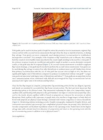
Figure 3. SIEV dissected 8 cm to length during DIEP flap elevation. DIEP: deep inferior epigastric perforator; SIEV: superficial inferior
epigastic vein.
Vein grafts can be used to increase pedicle length for either the arterial or venous anastomosis, augment flap
venous outflow with a second venous anastomosis through either the deep or superficial system, or during
flap salvage. Vein grafts are more commonly used during primary reconstruction to troubleshoot
intraoperative concerns . For example, if the recipient or flap vessels have scar or fibrosis, then cutting
[40]
back the vessels to more healthy tissue may shorten the vessel length resulting in the need for a vein graft. If
the primary recipient vessels are insufficient and pedicle length is needed to access alternative recipient
vessels, a vein graft may also be required [Figure 1]. If a second venous anastomosis is needed to augment
flap venous drainage, a vein graft may be needed to perform the second venous anastomosis. Vein grafts can
be used during flap salvage to restore flow after thrombosis or avulsion injury as well. Studies have shown
that primary reconstruction with the use of vein graft has success rates as high as 95%, but there are
significantly higher rates of thrombosis compared to primary reconstruction without vein graft . Longer
[44]
vein grafts are associated with higher rates of thrombosis and failure . Vein grafts are an important tool in
[45]
one’s armamentarium when confronted with difficult microsurgical free flap reconstructions but are
associated with higher rates of thrombosis and flap failure.
Once the free flap surgery is complete, postoperative flap monitoring protocols that are diligent, rigorous,
and timely are essential for successful free flap breast reconstruction. The first and most important flap
monitoring system is the physical exam. Flap assessment evaluating the skin color, temperature, turgor,
capillary refill, and bleeding with pinprick is the gold standard. Any signs of concern such as pale flap color,
cool to the touch, absent capillary refill or blood on pinprick are concerns for an arterial or inflow
thrombosis. Brisk capillary refill, hyperemic or purple skin color, swollen or boggy turgor, or brisk dark red
blood with pinprick are physical exam findings consistent with venous congestion or venous thrombosis
[Figure 4]. Monitoring systems including acoustic Doppler sonography, implantable Doppler devices, and
continuous near-infrared spectroscopy that complement physical exam can prompt early detection of
vascular compromise and facilitate expeditious return to the operating room. Studies have shown higher
salvage rates with a shorter time from detection of compromise and return to the operating room with the
[46]
use of a monitoring system in addition to serial physical exams .
Anticoagulation: