
Page 43 - Read Online
P. 43
Inchauste. Plast Aesthet Res 2023;10:27 https://dx.doi.org/10.20517/2347-9264.2022.139 Page 7 of 15
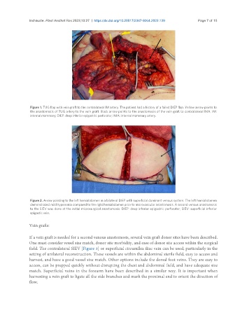
Figure 1. TUG flap with vein graft to the contralateral IM artery. The patient had a history of a failed DIEP flap. Yellow arrow points to
the anastomosis of TUG artery to the vein graft. Black arrow points to the anastomosis of the vein graft to contralateral IMA. IM:
internal mammary; DIEP: deep inferior epigastric perforator; IMA: internal mammary artery.
Figure 2. Arrow pointing to the left hemiabdomen in a bilateral DIEP with superficial dominant venous system. The left hemiabdomen
demonstrated mild hyperemia compared to the right hemiabdomen prior to microvascular anastomosis. A second venous anastomosis
to the SIEV was done at the initial microsurgical anastomosis. DIEP: deep inferior epigastric perforator; SIEV: superficial inferior
epigastic vein.
Vein grafts:
If a vein graft is needed for a second venous anastomosis, several vein graft donor sites have been described.
One must consider vessel size match, donor site morbidity, and ease of donor site access within the surgical
field. The contralateral SIEV [Figure 3] or superficial circumflex iliac vein can be used, particularly in the
setting of unilateral reconstruction. These vessels are within the abdominal sterile field, easy to access and
harvest, and have a good vessel size match. Other options include the dorsal foot veins. They are easy to
access, can be prepped quickly without disrupting the chest and abdominal field, and have adequate size
match. Superficial veins in the forearm have been described in a similar way. It is important when
harvesting a vein graft to ligate all the side branches and mark the proximal end to orient the direction of
flow.