
Page 19 - Read Online
P. 19
Chen et al. Plast Aesthet Res 2023;10:24 https://dx.doi.org/10.20517/2347-9264.2022.136 Page 9 of 26
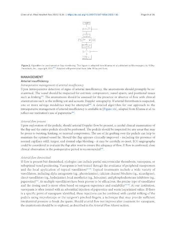
Figure 2. Algorithm for postoperative flap monitoring. This figure is adapted from Khansa et al. published in Microsurgery by Wiley
Periodicals, Inc., copyright 2013 [90] . Adapted with permission from John Wiley and Sons.
MANAGEMENT
Arterial insufficiency
Intraoperative management of arterial insufficiency
Upon intraoperative detection of signs of arterial insufficiency, the anastomosis should promptly be re-
examined. The vessel should be inspected for extrinsic compression, vessel spasm, and positional issues
such as kinking . The anastomosis should be assessed for the presence or absence of flow with clinical
[91]
examinations such as the milking test and acoustic Doppler sonography. If arterial thrombosis is suspected,
one or more salvage modalities may be attempted . A detailed algorithm for our approach to the
[90]
intraoperative management of arterial insufficiency is available in [Figure 3A], adapted from Khansa et al. to
reflect our institution’s use of papaverine .
[90]
Arterial flow present
Upon exploration of the pedicle, should arterial Doppler flow be present, a careful clinical examination of
the flap and the entire pedicle should be performed. The pedicle should be inspected for any areas that may
be prone to twisting, kinking, or external compression. The use of fat grafting over the pedicle can help to
maintain the optimal vessel lie. Should the flap appears clinically improved - including the presence of
normal capillary refill, turgor, and dermal edge bleeding - it may be carefully re-inset. ICG angiography
could be considered to evaluate the flap after inset to ensure the adequacy of flow. If flow is confirmed, close
clinical observation in the postoperative period is recommended .
[90]
Arterial flow diminished
If flow is present but diminished, etiologies can include partial microvascular thrombosis, vasospasm, or
suboptimal vessel positioning. Vasospasm is best treated through the avoidance of peripheral vasopressors
and the local application of topical vasodilators [92-95] . Topical treatments include a wide variety of
vasodilators, including alpha antagonists (eg., phentolamine), calcium channel blockers (eg., nicardipine),
direct vasodilators (eg., hydralazine), local anesthetics (eg., lidocaine), and phosphodiesterase inhibitors (eg.,
[96]
papaverine) . As multiple vasodilators have been proven to be efficacious, the precise type of vasodilator
and the dosing used is more often based on surgeon experience and availability [95-98] . At our institution,
vasospasm is often treated with an adventitial injection of papaverine and warm heparinized saline. If there
is a specific point of vasospasm identified, these injections can be combined with careful milking of the
pedicle using microforceps or the surgeon’s pinched fingers, a technique that may provide sufficient
intraluminal pressure to break the spasm. Should arterial flow not improve after treatment for vasospasm,
the anastomosis should be re-explored, as described in the Arterial Flow Absent section.