
Page 22 - Read Online
P. 22
Page 12 of 26 Chen et al. Plast Aesthet Res 2023;10:24 https://dx.doi.org/10.20517/2347-9264.2022.136
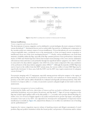
Figure 4. Algorithm for postoperative treatment of microvascular thrombosis.
Venous insufficiency
Venous congestion and venous thrombosis
The development of venous congestion can be attributed to several etiologies, the most common of which is
[104]
venous thrombosis . Mechanical factors such as unfavorable flap position, or kinking and compression of
the vascular pedicle are common causes of venous insufficiency. Venous thrombosis can occur secondary to
a hypercoagulable state, a technical error at the anastomosis, or from prolonged venous congestion or
insufficiency from one of the above mechanical factors [105,106] . Finally, venous insufficiency can result from
anatomic variability within the flap, especially if the flap contains portions of two perforasomes. Anatomical
studies of DIEP and TRAM flaps have shown that normal venous drainage of the lower anterior abdominal
subcutaneous tissue and skin occurs primarily through the superficial inferior epigastric vein (SIEV), which
is connected to the deep inferior epigastric vein (DIEV) by choke vessels composed of the vena comitantes
[107]
of the perforators of the deep inferior epigastric artery (DIEA) . Although the majority of DIEP flaps may
survive based on the outflow from the DIEV system alone, the venous outflow of some DIEP flaps may be
superficially dominant. In these cases, both the DIEV and the SIEV may require anastomosis for adequate
venous drainage .
[107]
Preoperative imaging with CT angiogram, especially among patients with prior surgery in the region of
planned flap harvest, may be beneficial in perforator selection and evaluation of venous anatomy. The
presence of the SIEV and its caliber should also be evaluated radiologically and intraoperatively. If the SIEV
is noted to be of good caliber (> 1.5 mm), it is prudent to preserve adequate length on this vessel to allow for
anastomosis.
Intraoperative management of venous insufficiency
As described by Heller and Levin, obstruction of venous outflow can lead to red blood cell extravasation,
endothelial breakdown, microcirculation thrombosis, and flap death . Signs of venous congestion in the
[108]
flap may include rapid capillary refill on the skin paddle (< 1-2 seconds), more profuse dermal bleeding of a
darker color, loss of venous Doppler signals in the pedicle and perforators, greater flap turgor, and enlarged
secondary veins such as the SIEV. Our algorithm for intraoperative management of suspected venous
insufficiency is available in [Figure 3B], adapted from Khansa et al. to reflect our institution’s use of leeching
[90]
and de-epithelialization .
Suspicion for venous congestion requires release of insetting sutures and diligent assessment of vessel
position, flap and pedicle orientation within the breast pocket, and presence of hematoma and/or edema