
Page 131 - Read Online
P. 131
Foppiani et al. Plast Aesthet Res 2023;10:53 https://dx.doi.org/10.20517/2347-9264.2022.137 Page 5 of 14
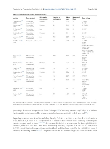
Table 1. Study characteristics and flap demographics
NIH quality Number of Mean Number of
Author Type of study Type of flap
assessment participants age flaps
[19]
Thiessen et al. Prospective Moderate 21 56.7 33 1 TRAM
2020 observational 32 DIEP
[20]
Saxena et al. Prospective Moderate 32 51.9 32 32 TRAM
2019 observational
[21]
Phillips et al. Prospective Low 19 54.6 30 30 DIEP
2020 observational
Lindelauf et al. [22] Prospective Moderate 30 51 42 42 DIEP
2021 observational
Johnson et al. [23] Retrospective Low 460 50.7 740 740 “abdominal-based
2021 observational flaps”
Pelletier et al. [24] Randomized control Low 50 49.2 50 14 TRAM
2011 21 DIEP
9 SIEA
3 DIEP/SIEA double
stacked flaps
3 DIEP/SIEV
turbocharged flaps
1 DIEP + DIEP double
stacked flap
Ricci et al. [25] Retrospective Low 900 50.3 900 3 TRAM
2017 observational 872 DIEP
2 SIEA
23 SGAP
Ozturk et al. [26] Prospective Moderate 20 49.3 30 4 TRAM
2014 observational 24 DIEP
2 SIEA
[27]
Saad et al. Retrospective Moderate 120 53 120 35 TRAM
2020 observational 85 DIEP
[28]
Salgarello et al. Retrospective Moderate 45 52.6 45 45 DIEP
2018 observational
Carruthers et al. [29] Retrospective Low 196 50.7 301 301 DIEP
2019 observational
Tran et al. [30] Retrospective Low 175 50.9 286 3 MS-TRAM
2021 observational 266 DIEP
3 SIEA
6 TUG/DUG
8 PAP
[31]
Kumbasar et al. Prospective High 10 57 10 1 TRAM
2021 observational 8 DIEP
1 LD
[32]
Koolen et al. Retrospective Low 451 48.9 670 3 TRAM
2016 observational 646 DIEP
1 SIEA
20 SGAP
NIH: National Institute of Health; DIEP: deep inferior epigastric; TRAM: transverse rectus abdominis; SGAP: superior gluteal artery perforator;
SIEA: superficial inferior epigastric artery; PAP: profunda artery perforator; DUG/TUG: diagonal/transverse upper gracilis; LD: latissimus dorsi.
providing a short-term perspective on thermal changes [19,20] . Conversely, the study by Phillips et al. did not
[21]
furnish details on their protocol for measurements, leaving some ambiguity in their approach .
Regarding oximetry, several studies-including those by Pelletier et al., Ricci et al., Ozturk et al., Carruthers
et al., Tran et al., Koolen et al., and Johnson et al.-relied on the ViOptix tissue oximetry technology to
monitor oxygen levels in tissue [23-26,29,30,32] . In contrast, Lindelauf et al. employed the Foresight MC-2030
oximeter, Saad et al. used the T-Stat tissue oximeter by Spectros, Salgarello et al. utilized the Somanetics
INVOS 5,100 C Cerebral/Somatic Oximeter (Covidien), and Kumbasar opted for the INVOS 700 cerebral
oximetry monitoring system [22,27,28,31] . The protocols for the use of these diagnostic tools exhibited some