
Page 108 - Read Online
P. 108
Page 286 Cencioni. Neuroimmunol Neuroinflammation 2020;7:277-90 I http://dx.doi.org/10.20517/2347-8659.2020.18
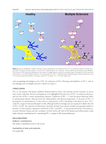
Figure 2. Immune checkpoints inhibitor treatment induces reactivation of multiple Sclerosis. PD-1 has been described on autoreactive T
cells and TFH cells in multiple sclerosis. The link of PD-1 with the ligand PDL-1 controls T cell activation mantaining tollerance (Healthy).
The blocking of PD-1 breaks the tollerance by reactivation of antigen-specific cells and causing damage of the target-cells with persistent
+
inflammation in the CNS (Multiple Sclerosis). Contrary, depletion of PD-1 T cells eliminates activated antigen-specific cells reducing the
T cell infiltrates and inflammation in the target-organ (PD-1 blocking). TFH: T Follicular helper cells
+
cells recognising self-antigen in the CNS. The relevance of PD-1 blocking and depletion of PD-1 cells in
the pathogenesis of multiple sclerosis is shown in Figure 2.
CONCLUSION
PD-1 is an immune checkpoint inhibitor demonstrated to reduce the immune system response in cancer
and chronic infection. Recent investigations have highlighted the dual role of PD-1 in immune tolerance,
+
and the loss of PD-1 causes autoimmune diseases. Depletion of PD-1 T cells has given beneficial effect
in autoimmune disease, slowing down the inflammation and disease progression. A decrease of PD-1 is
predisposed to autoimmunity, as described in experiments of PD-1 blocking or knockout in mice. PD-1
could be a target of immunotherapies in MS, although further investigations are required to define the role
of PD-1 in MS. The majority of information has been derived from animal models and sporadic studies in
humans. To this purpose, expression and levels of PD-1 and PDL-1 in the peripheral blood, CSF and post-
mortem MS brain tissues and the correlation of their levels with risk of MS disease and inflammation could
make relevant contributions for considering PD-1 a target in MS immunotherapies.
DECLARATIONS
Authors’ contributions
The author contributed solely to the article.
Availability of data and materials
Not applicable.