
Page 77 - Read Online
P. 77
Das et al. Neuroimmunol Neuroinflammation 2020;7:141-9 I http://dx.doi.org/10.20517/2347-8659.2020.36 Page 145
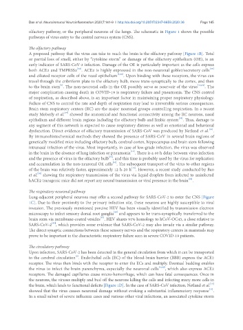
olfactory pathway, or the peripheral neurons of the lungs. The schematic in Figure 1 shows the possible
pathways of virus entry to the central nervous system (CNS).
The olfactory pathway
A proposed pathway that the virus can take to reach the brain is the olfactory pathway [Figure 1B]. Total
or partial loss of smell, either by “cytokine storm” or damage of the olfactory epithelium (OE), is an
early indicator of SARS-CoV-2 infection. Damage of the OE is particularly important as the cells express
[35]
[34]
both ACE2 and TMPRSS2 . ACE2 is highly expressed in the non-neuronal goblet/secretory cells
and ciliated receptor cells of the nasal epithelium [5,36] . Upon binding with these receptors, the virus can
travel through the cribriform plate to the olfactory bulb, move trans-synaptically to the cortex, and then
[5]
to the brain stem . The non-neuronal cells in the OE possibly serve as reservoir of the virus [13,34] . The
major complication causing death in COVID-19 is respiratory failure and pneumonia. The CNS control
of respiration, as described above, is an important factor in maintaining proper respiratory physiology.
Failure of CNS to control the rate and depth of respiration may lead to irreversible serious consequences.
Brain stem respiratory centers (RC) are the major neuronal groups controlling respiration. In a recent
[37]
study Moberly et al. showed the anatomical and functional connectivity among the RC neurons, nasal
[37]
epithelium and different brain regions including the olfactory bulb and limbic system . Thus, damage to
any segment of this network is expected to cause respiratory distress as well as emotional and behavioral
[13]
dysfunction. Direct evidence of olfactory transmission of SARS-CoV was produced by Netland et al. .
By immunohistochemical methods they showed the presence of SARS-CoV in several brain regions of
genetically modified mice including olfactory bulb, cerebral cortex, hippocampus and brain stem following
intranasal infection of the virus. Most importantly, in case of low-grade infection, the virus was observed
[13]
in the brain in the absence of lung infection or pneumonia . There is a 60 h delay between virus infection
[13]
and the presence of virus in the olfactory bulb , and this time is probably used by the virus for replication
[34]
and accumulation in the non-neuronal OE cells . The subsequent transport of the virus to other regions
[13]
of the brain was relatively faster, approximately 12 h-20 h . However, a recent study conducted by Bao
[38]
et al. showing the respiratory transmission of the virus via liquid droplets from infected to uninfected
hACE2 transgenic mice did not report any neural transmission or viral presence in the brain .
[38]
The respiratory neuronal pathway
Lung-adjacent peripheral neurons may offer a second pathway for SARS-CoV-2 to enter the CNS [Figure
1C]. Due to their proximity to the primary infection site, these neurons are highly susceptible to viral
invasion. The previously mentioned porcine HEV has been visually identified by transmission electron
[39]
microscopy to infect sensory dorsal root ganglia and appears to be trans-synaptically transferred to the
[40]
brain stem via membrane-coated vesicles . HEV shares 91% homology to hCoV-OC43, a close relative to
[41]
SARS-CoV-2 , which provides some evidence that SARS-CoV-2 may also invade via a similar pathway.
The direct synaptic connections between these sensory nerves and the respiratory centers in mammals may
prove to be important in the characteristic respiratory failure seen in severe COVID-19 patients.
The circulatory pathway
Upon infection, SARS-CoV-2 has been detected in the general circulation from which it can be transported
to the cerebral circulation . Endothelial cells (EC) of the blood-brain barrier (BBB) express the ACE2
[5]
receptor. The virus then binds with the receptor to enter the ECs and multiply. Eventual budding enables
the virus to infect the brain parenchyma, especially the neuronal cells [5,42] , which also express ACE2
receptors. The damaged capillaries cause micro-hemorrhage, which can have fatal consequences. Once in
the neurons, the viruses multiply and bud off the neurons killing the cells and infecting many more cells in
[13]
the brain, which leads to functional deficits [Figure 1D]. In the case of SARS-CoV infection, Netland et al.
showed that the virus causes neuronal damage without evoking a substantial inflammatory response .
[13]
In a small subset of severe influenza cases and various other viral infections, an associated cytokine storm