
Page 75 - Read Online
P. 75
Das et al. Neuroimmunol Neuroinflammation 2020;7:141-9 I http://dx.doi.org/10.20517/2347-8659.2020.36 Page 143
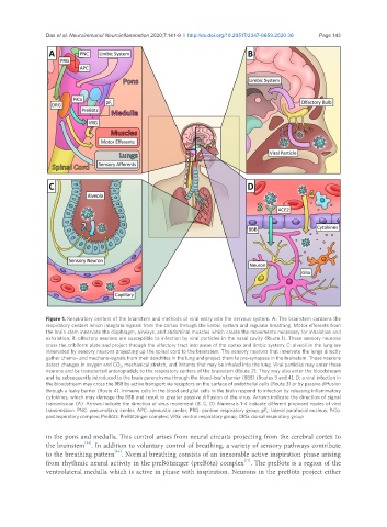
Figure 1. Respiratory centers of the brainstem and methods of viral entry into the nervous system. A: The brainstem contains the
respiratory centers which integrate signals from the cortex through the limbic system and regulate breathing. Motor efferents from
the brain stem innervate the diaphragm, airways, and abdominal muscles which create the movements necessary for inhalation and
exhalation; B: olfactory neurons are susceptible to infection by viral particles in the nasal cavity (Route 1). These sensory neurons
cross the cribiform plate and project through the olfactory tract into areas of the cortex and limbic system; C: alveoli in the lung are
innervated by sensory neurons projecting up the spinal cord to the brainstem. The sensory neurons that innervate the lungs directly
gather chemo- and mechano-signals from their dendrites in the lung and project them to pre-synapses in the brainstem. These neurons
detect changes in oxygen and CO 2 , mechanical stretch, and irritants that may be inhaled into the lung. Viral particles may enter these
neurons and be transported anterogradely to the respiratory centers of the brainstem (Route 2). They may also enter the bloodstream
and be subsequently introduced to the brain parenchyma through the blood–brain barrier (BBB) (Routes 3 and 4); D: a viral infection in
the bloodstream may cross the BBB by active transport via receptors on the surface of endothelial cells (Route 3) or by passive diffusion
through a leaky barrier (Route 4). Immune cells in the blood and glial cells in the brain respond to infection by releasing inflammatory
cytokines, which may damage the BBB and result in greater passive diffusion of the virus. Arrows indicate the direction of signal
transmission (A). Arrows indicate the direction of virus movement (B, C, D). Numerals 1-4 indicate different proposed routes of viral
transmission. PNC: pneumotaxic center; APC: apneustic center; PRG: pontine respiratory group; pF L : lateral parafacial nucleus; PiCo:
postinspiratory complex; PreBötz: PreBötzinger complex; VRG: ventral respiratory group; DRG: dorsal respiratory group
in the pons and medulla. This control arises from neural circuits projecting from the cerebral cortex to
[15]
the brainstem . In addition to voluntary control of breathing, a variety of sensory pathways contribute
[16]
to the breathing pattern . Normal breathing consists of an inexorable active inspiration phase arising
from rhythmic neural activity in the preBötzinger (preBötz) complex . The preBötz is a region of the
[17]
ventrolateral medulla which is active in phase with inspiration. Neurons in the preBötz project either