
Page 65 - Read Online
P. 65
Griffiths et al. Neuroimmunol Neuroinflammation 2020;7:51-67 I http://dx.doi.org/10.20517/2347-8659.2019.21 Page 61
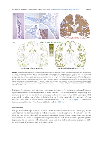
Figure 5. Meningeal inflammation and subpial cortical demyelination. Example infiltrates of the leptomeninges overlying the neocortex
in a single coronal macrosection. Aggregates visualised by LFB/haematoxylin histology were rich in CD20+ B cells (A). The location
of the infiltrates in (A) are marked as asterisks (*) on the serial anti-PLP stained section (B) (Case MS217). By grouping the cases
based on their relative extent of leptomeningeal inflammation (0/+ vs. ++/+++), we constructed schema depicting all neocortical grey
matter lesions noted per P1 section, per case (n = 18). Depth of colour (see heatmap representing percent lesion occurrence with 100%
indicating tissue was demyelinated in every P1 section in that group) illustrates how frequently an area of neocortex was demyelinated
in meningeal inflammation rated 0/+ and meningeal inflammation rated ++/+++ MS (C). PLP: proteolipid protein. (A) Scale bar = 200
µm; (B) Scale bar = 1 cm
lesion area (10.1%, range 0.6%-42.1% vs. 10.0%, range 2.3%-26.5%, P = 0.981) was unchanged between
leptomeningeal rated moderate-high cases vs. those rated with little-to-mild infiltrates, respectively. The
relationship between the extent of leptomeningeal inflammation and cortical GML area is presented
schematically as a heat map of all cortical GMLs observed (in olive green colour) in section P1 of cases
with mild (rated 0 to +, n = 9) vs. modest/substantial infiltrates (++ to +++, n = 9; Figure 5C). Please note
that four cases did not have P1 sections available for analysis [Table 1].
DISCUSSION
Our quantitative histological analysis of whole coronal macrosections demonstrates cortical grey matter
demyelination can be the preeminent pathology in some cases of progressive MS and can be used to
identify a post-mortem cohort with a more severe pathological disease. Subpial cortical grey matter lesions
associated with the extent of demyelinated deep grey matter and with infiltrates of the leptomeninges but
importantly did not associate with white matter lesion load. Our study reinforces the concept of a CSF-
derived (outside-in) driver of tissue pathology in SPMS.