
Page 98 - Read Online
P. 98
Page 20 of 32 Noor et al. Neuroimmunol Neuroinflammation 2019;6:10 I http://dx.doi.org/10.20517/2347-8659.2019.18
A B
C D
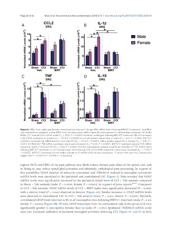
Figure 6. DRGs from males and females reveal similarly reduced IL-1β and TNF mRNA levels following BIRT377 treatment. Total RNA
was isolated from ipsilateral lumbar DRGs from the same mice used in Figure 2B, and analyzed for inflammatory cytokines. (A) In the
DRGs, CCI induced CCL2 mRNA levels (F 1,1 = 25.5, P < 0.0001) remained unchanged following BIRT377 treatment. (B) CCI increased
IL-1β mRNA expression in females (F 1,1 = 25.4, P < 0.0001) to a much greater degree than in males (F 1,1 = 8.3, P = 0.006). BIRT377
treatment reduced IL-1β mRNA levels in mice with CCI (F 1,1 = 25.4, P < 0.0001), with a greater magnitude in females (F 1,1 = 4.95, P =
0.031). (C) Post-CCI TNF mRNA expression levels were increased (F 1,1 = 61.81, P < 0.0001). BIRT377 treatment reduced TNF mRNA
expression levels in mice with CCI (F 1,1 = 9.92, P = 0.003). Post hoc comparisons revealed a significant reduction of TNF mRNA levels
following BIRT377 treatment in CCI-treated males. (D) Following CCI, IL-10 mRNA expression levels were increased (F 1,1 = 77.99, P
< 0.0001). BIRT377 treatment did not further elevate IL-10 mRNA levels during neuropathy. *P values from post hoc comparisons
ranges from P = 0.029 to P < 0.0001, n = 6 per group
regions (SCN and DRG) of the pain pathway may likely reduce chronic pain relays to the spinal cord, and
in doing so, may reduce spinal glial activation and ultimately, pathological pain processing. In support of
this possibility, GFAP (marker of astrocyte activation) and TMEM119 (related to microglial activation)
mRNA levels were examined in the ipsilateral and contralateral LSC [Figure 8]. Data revealed that GFAP
mRNA levels were significantly increased in the ipsilateral dorsal horn of CCI + Veh animals compared
to Sham + Veh animals (male: P = 0.0001; female: P < 0.0001), in support of prior reports [26,84] . Compared
to CCI + Veh animals, GFAP mRNA levels of CCI + BIRT males were significantly decreased (P = 0.044),
with a similar trend (P = 0.056) observed in females [Figure 8A]. Similar increases in GFAP mRNA levels
were observed in contralateral LSC in CCI + Veh animals (male: P = 0.015; female: P < 0.0001). Similarly,
contralateral GFAP levels returned to BL in all neuropathic mice following BIRT377 treatment (male: P = 0.04;
female: P = 0.0003) [Figure 8B]. Of note, GFAP transcripts from the contralateral side of the spinal cord were
significantly greater in neuropathic females than in males (P = 0.001). Ipsilateral TMEM119 mRNA levels
were also increased, indicative of increased microglial activation following CCI [Figure 8C and D] in both