
Page 46 - Read Online
P. 46
Page 2 of 4 Orsucci et al. Neuroimmunol Neuroinflammation 2018;5:6 I http://dx.doi.org/10.20517/2347-8659.2017.66
on the left side of the body. Both the family history and past medical history were unremarkable. At
that time, brain magnetic resonance imaging (MRI) was normal except few non-specific white matter
lesions. Electroencephalography (EEG) (including prolonged monitoring) was normal. He was prescribed
acetylsalicylic acid (ASA) 100 mg/day, but chose to discontinue using after an episode of nosebleeding.
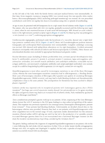
At age 40 years, he presented with left hemiparesis due to a right mesial frontal ischemic stroke [Figure 1A and B].
The patient, who was prescribed with ASA 300 mg/day, gradually recovered until the following year (aged
41 years), when he was examined because of acute-onset left hemianopia. MRI showed an acute ischemic
lesion in the right temporo-parietal-occipital regions [Figure 1C and D]. Ivy bleeding time was prolonged to
[1]
14 min (normal 2.5-11 min ) confirming appropriate antiaggregation.
Cerebrovascular angiography, performed under the hypothesis of a vasculitis, showed only a right fetal-
type posterior cerebral artery (PCA) [Figure 1E and F]. Brain 18F-fluorodeoxyglucose positron emission
tomography and cerebrospinal fluid examination were unremarkable. Complete cardiologic screening
was normal. EEG showed mild epileptiform alterations on the right hemisphere. Cerebral autosomal
dominant arteriopathy with subcortical infarcts and leukoencephalopathy (CADASIL), Fabry disease and
mitochondrial disorders were excluded by appropriate biochemical and genetic studies.
Routine laboratory assays (including D-dimer, prothrombin time, activated partial thromboplastin time,
factor V, antithrombin, protein C, protein S, activated protein C resistance, lupus anticoagulans, anti-
antinuclear antibodies, anti-smooth muscle antibodies, anti-cardiolipin antibodies, extractable nuclear
antigen antibodies, anti-neutrophil cytoplasmic antibodies and antiphospholipid antibodies) were normal,
except for a mild but long-lasting hypofibrinogenemia (115-180 mg/dL; normal 200-400 mg/dL).
Hypofibrinogenemia is most often caused by heterozygous mutations in one of the three fibrinogen
chains, whereas the same homozygous mutations commonly lead to afibrinogenemia, a bleeding disease.
Most cases of heterozygous disorders of fibrinogen affect quantity and quality of circulating fibrinogen
(hypodysfibrinogenemia, OMIM #616004), and may lead to both bleeding diathesis and thromboembolic
complications (even in the same patient). This predisposition for thrombosis has been ascribed to delayed
[2]
plasmin digestion .
Ischemic stroke was reported only in exceptional patients with homozygous gamma chain (FGG)
[3]
mutations , but large case-control association studies showed that polymorphisms in the genes encoding
the alpha and gamma chains may lead to an increased risk of ischemic stroke, likely mediated by qualitative
[4]
effects on fibrinogen (and fibrin) .
These considerations prompted a genetic work-up in our patients. At last, sequencing of the three fibrinogen
chains showed the 945C>T mutation in the FGG gene, leading to a stop codon (p.Arg108Stop of the mature
chain). This mutation was previously reported in ten consanguineous, homozygous afibrinogenemic patients
[5]
from Lebanon . These patients had bleeding diathesis. Unfortunately, no detailed clinical data on the
heterozygous relatives were provided. However they had, as our patients, mild hypofibrinogenemia with
other coagulation tests in the normal range. Of note, expression studies demonstrated that this nonsense
mutation affected neither mRNA splicing nor stability, but led to the production of an unstable, severely
truncated fibrinogen gamma chain that is not incorporated into a functional fibrinogen hexamer (in this
[5]
study the mutation was denoted as p.Arg134Stop, considering the immature chain) .
The genetic finding confirmed the diagnosis of congenital hypodysfibrinogenemia in our patient, who in the
3 years following the 2nd stroke did not experience further TIAs or stroke (under double-antiaggregation
therapy with ASA 100 mg/day and clopidogrel 75 mg/day). He had only 3 episodes of simple motor partial
seizures successfully treated with oxcarbazepine.