
Page 42 - Read Online
P. 42
Page 8 of 10 Dong et al. Neuroimmunol Neuroinflammation 2018;5:5 I http://dx.doi.org/10.20517/2347-8659.2017.47
Exosomes or exocrine secretion
Caused spreading of pathological TDP-43
Autophagosomes
Exosomes
TDP-43 pathological deposition
TDP-43 aggregation Caused axonal swelling and
impaired the mobility
Proteasome
TDP43 25-kDa
TDP-43 protein Nuclear Intracytoplasmic inclusion
TARDBP mutation caused
mis-localization
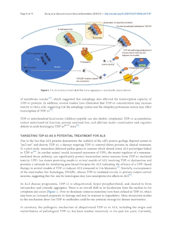
Figure 1. The role of ubiquitinated tdp43 that forms aggregates in amyotrophic lateral sclerosis
of membrane vesicles , which suggested that autophagy also affected the transcription capacity of
[26]
TDP-43 proteins. In addition, several studies have illustrated that TDP-43 concentration may increase
toxicity in HeLa cells, suggesting that the autophagy system and the ubiquitin proteasome system may affect
[34]
transcription of TDP-43 .
TDP-43 mitochondrial localization inhibitory peptide can also abolish cytoplasmic TDP-43 accumulation,
restore mitochondrial function, prevent neuronal loss, and alleviate motor-coordinative and cognitive
[35]
deficits in adult hemizygous TDP-43 M337V mice .
TARGETING TDP-43 AS A POTENTIAL TREATMENT FOR ALS
Due to the fact that ALS patients demonstrate the inability of the cell’s protein garbage disposal system to
“pull out” and destroy TDP-43, a therapy targeting TDP-43 removal shows promise in clinical treatment.
In a pilot study, researchers delivered parkin genes to neurons which slowed down ALS pathologies linked
[36]
to TDP-43 . In another animal model, increased expression of UPF1, the master regulator of a nonsense-
mediated decay pathway, can significantly protect mammalian motor neurons from TDP-43 mediated
toxicity. UPF1 has shown promising results in animal models of ALS involving TDP-43 dysfunction and
provides a rationale for developing gene-based therapies for ALS indicating the efficacy of a UPF1-based
[37]
therapy in animal models of TDP-43 induced ALS pioneered in this laboratory . Similarly, overexpression
of the mammalian Sis1 homologue, DNAJB1, relieves TDP-43 mediated toxicity in primary rodent cortical
[38]
neurons, suggesting that Sis1 and its homologues may have neuroprotective effects in ALS .
In ALS disease progression, TDP-43 is ubiquitinated, hyper-phosphorylated, and cleaved to form
intranuclear and cytosolic aggregates. There is an overall shift in its localization from the nucleus to the
cytoplasm and axons [Figure 1]. Over 60 dominant missense mutations have been defined in TDP-43, which
may have an increased propensity to cleavage and may be resistant to degradation. More stimulation studies
in this mechanism show that TDP-43 antibodies could be one potential strategy for disease intervention.
In summary, the pathogenic mechanism of ubiquitinated TDP-43 in ALS, including the origin and
redistribution of pathological TDP-43, has been studied intensively in the past ten years. Currently,