
Page 408 - Read Online
P. 408
Page 4 of 6 Almedallah et al. Neuroimmunol Neuroinflammation 2018;5:48 I http://dx.doi.org/10.20517/2347-8659.2018.55
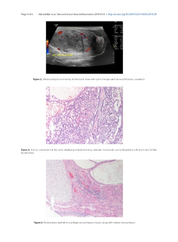
Figure 2. Ultrasounds picture showing left testicular mass with cystic changes and microcalcifications, neoplastic
Figure 3. Yolk sac component of the tumor displaying endodermal sinus, reticular, microcystic, and solid patterns with prominent Schiller
Duval bodies
Figure 4. Teratomatous epithelium (cartilage and pulmonary tissue), along with mature nervous tissue