
Page 413 - Read Online
P. 413
Paraskevas et al. Neuroimmunol Neuroinflammation 2018;5:49 I http://dx.doi.org/10.20517/2347-8659.2018.50 Page 3 of 6
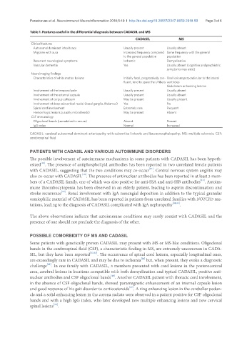
Table 1. Features useful in the differential diagnosis between CADASIL and MS
CADASIL MS
Clinical features
Autosomal dominant inheritence Usually present Usually absent
Migraine with aura Increased frequency compared Same frequency with the general
to the general population population
Recurrent neurological symptoms Ischemic Demyelinative
Vascular dementia Yes Usually absent (cognitive and psychiatric
symptoms may exist)
Neuroimaging findings
Characteristics of white matter lesions Initially focal, progressively con- Oval lesions perpendicular to the lateral
fluent, tend to spare the U fibers ventricles
Gadolinium enhancing lesions
Involvement of the temporal pole Usually present Usually absent
Involvement of the external capsule Usually present Usually absent
Involvement of corpus callosum May be present Usually present
Involvement of deep subcortical nuclei (basal ganglia, thalamus) Yes No
Spinal cord involvement Extremely rare Frequent
Hemorrhagic lesions (usually microbleeds) May be present Absent
CSF immunology
Oligoclonal bands (unmatched in serum) Absent Present
IgG index Normal Increased
CADASIL: cerebral autosomal dominant arteriopathy with subcortical infarcts and leucoencephalopathy; MS: multiple sclerosis; CSF:
cerebrospinal fluid
PATIENTS WITH CADASIL AND VARIOUS AUTOIMMUNE DISORDERS
The possible involvement of autoimmune mechanisms in some patients with CADASIL has been hypoth-
[16]
esized . The presence of antiphospholipid antibodies has been reported in two unrelated female patients
[17]
with CADASIL, suggesting that the two conditions may co-occur . Central nervous system angiitis may
[18]
also co-occur with CADASIL . The presence of antinuclear antibodies has been reported in at least 3 mem-
[11]
bers of a CADASIL family, one of which was also positive for anti-SSA and anti-SSB antibodies . Autoim-
mune thrombocytopenia has been observed in an elderly patient, leading to aspirin discontinuation and
[19]
stroke recurrence . Renal involvement with IgA mesangial deposition in addition to the typical granular
osmiophilic material of CADASIL has been reported in patients from unrelated families with NOTCH3 mu-
tations, leading to the diagnosis of CADASIL complicated with IgA nephropathy [20,21] .
The above observations indicate that autoimmune conditions may rarely coexist with CADASIL and the
presence of one should not preclude the diagnosis of the other.
POSSIBLE COMORBIDITY OF MS AND CADASIL
Some patients with genetically proven CADASIL may present with MS or MS-like conditions. Oligoclonal
bands in the cerebrospinal fluid (CSF), a characteristic finding in MS, are extremely uncommon in CADA-
SIL, but they have been reported [22,23] . The occurrence of spinal cord lesions, especially longitudinal ones,
[24]
are exceedingly rare in CADASIL and may be due to ischemia but, when present, they evoke a diagnostic
[25]
challenge . In one family with CADASIL, 3 members presented with cord lesions in the posterocentral
area, cerebral lesions in locations compatible with both demyelination and typical CADASIL, positive anti-
[26]
nuclear antibodies and CSF oligoclonal bands . Another CADASIL patient with thoracic cord involvement,
in the absence of CSF oligoclonal bands, showed paramagnetic enhancement of an internal capsule lesion
[27]
and good response of his gait disorder to corticosteroids . A ring enhancing lesion in the cerebellar pedun-
cle and a solid enhancing lesion in the corona radiate were observed in a patient positive for CSF oligoclonal
bands and with a high IgG index, who later developed new multiple enhancing lesions and new cervical
[28]
spinal lesions .