
Page 407 - Read Online
P. 407
Almedallah et al. Neuroimmunol Neuroinflammation 2018;5:48 I http://dx.doi.org/10.20517/2347-8659.2018.55 Page 3 of 6
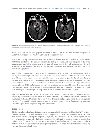
Figure 1. Fluid-attenuated inversion recovery magnetic resonance imaging of the brain showing hyperintensity within the medial aspect of
the temporal lobes suggestive of limbic encephalitis
aspartic acid (NMDA), anti-voltage-gated potassium channels (VGKC), anti-contactin associated protein 2
(CASPR2) and anti-LG1 was conducted, but all yielded negative results.
Due to his neurological state at the time, our patient was deemed an unfit candidate for chemotherapy,
and tumor resection was the mainstay approach for treating the cancer. Left radical inguinal orchiectomy
was done and revealed the mass to be a mixed germ cell tumor constituting yolk sac tumor 90% [Figure 3]
and teratoma 10% [Figure 4]. The resection was intended to treat the cancer itself and halt the source of the
neurological symptoms.
The oncology team decided against adjuvant chemotherapy after the resection, and were reassured by
the regression in lymph node sizes. His level of consciousness improved and his speech became more
clear. However, there was no improvement in his memory, orientation, or gaze. In the following days, he
returned to his baseline status upon presentation where he experienced excessive sleepiness and more
visual hallucinations. The patient underwent multiple sessions of pulse steroid therapy, intravenous
immunoglobulin (IVIG) and rituximab but there was no clinical improvement. Moreover, the patient started
to develop seizures after the initial IVIG session and was then switched to rituximab. The seizures were slow
with no epileptiform discharges, and despite the change in treatment they recurred frequently.
In the subsequent months, our patient continued to deteriorate in terms of level of consciousness and
respiratory function. He was admitted to the intensive care unit (ICU) where he later became dependent on
mechanical ventilation. During his stay in the ICU, he developed multiple septic shocks due to his impaired
immunity from tacrolimus as an attempt to prevent liver transplant rejection and unfortunately later went
into multi-organ failure. He passed away from cardiac arrest.
DISCUSSION
[3]
Limbic encephalitis has been first described as a paraneoplastic syndrome by Corsellis et al. in 1968.
Considered to be a rare association of common malignancies, such as small cell lung cancer, testicular
[4]
[5]
and ovarian cancers , Hodgkin’s lymphoma, thymoma, and teratomas . The incidence depends on the
type of associated cancer; with the lowest incidence of 8% attributed to testicular cancer, as described by
[1]
Gultekin et al. . This incidence is expected to increase with the development of new diagnostic methods
and treatment modalities, which will lead to prolonged survival of cancer patients and hence increase the
number of diagnosis of such cases.