
Page 32 - Read Online
P. 32
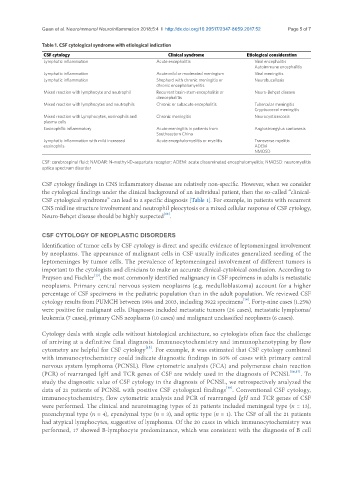
Guan et al. Neuroimmunol Neuroinflammation 2018;5:4  I  http://dx.doi.org/10.20517/2347-8659.2017.52                     Page 5 of 7


               Table 1. CSF cytological syndrome with etiological indication
                CSF cytology                              Clinical syndrome         Etiological consideration
                Lymphatic inflammation                Acute encephalitis               Viral encephalitis
                                                                                       Autoimmune encephalitis
                Lymphatic inflammation                Acute mild or moderated meningism  Viral meningitis
                Lymphatic inflammation                Shepherd with chronic meningitis or   Neurobucellosis
                                                      chronic encephalomyelitis
                Mixed reaction with lymphocyte and neutrophil  Recurrent brain-stem encephalitis or   Neuro-Behçet disease
                                                      diencephalitis
                Mixed reaction with lymphocytes and neutrophils  Chronic or subacute encephalitis   Tubercular meningitis
                                                                                       Cryptococcal meningitis
                Mixed reaction with Lymphocytes, eosinophils and   Chronic meningitis  Neurocysticercosis
                plasma cells
                Eosinophilic inflammatory             Acute meningitis in patients from   Angiostrongylus cantonesis
                                                      Southeastern China
                Lymphatic inflammation with mild increased   Acute encephalomyelitis or myelitis  Transverse myelitis
                eosinophils                                                            ADEM
                                                                                       NMOSD

               CSF: cerebrospinal fluid; NMDAR: N-methyl-D-aspartate receptor; ADEM: acute disseminated encephalomyelitis; NMOSD: neuromyelitis
               optica spectrum disorder

               CSF cytology findings in CNS inflammatory disease are relatively non-specific. However, when we consider
               the cytological findings under the clinical background of an individual patient, then the so-called “clinical-
               CSF cytological syndrome” can lead to a specific diagnosis [Table 1]. For example, in patients with recurrent
               CNS midline structure involvement and neutrophil pleocytosis or a mixed cellular response of CSF cytology,
                                                        [12]
               Neuro-Behçet disease should be highly suspected .


               CSF CYTOLOGY OF NEOPLASTIC DISORDERS
               Identification of tumor cells by CSF cytology is direct and specific evidence of leptomeningeal involvement
               by neoplasms. The appearance of malignant cells in CSF usually indicates generalized seeding of the
               leptomeninges by tumor cells. The prevalence of leptomeningeal involvement of different tumors is
               important to the cytologists and clinicians to make an accurate clinical-cytoloical conclusion. According to
                                 [13]
               Prayson and Fischler , the most commonly identified malignancy in CSF specimens in adults is metastatic
               neoplasms. Primary central nervous system neoplasms (e.g. medulloblastoma) account for a higher
               percentage of CSF specimens in the pediatric population than in the adult population. We reviewed CSF
                                                                                   [14]
               cytology results from PUMCH between 1984 and 2003, including 3922 specimens . Forty-nine cases (1.25%)
               were positive for malignant cells. Diagnoses included metastatic tumors (26 cases), metastatic lymphoma/
               leukemia (7 cases), primary CNS neoplasms (10 cases) and malignant unclassified neoplasms (6 cases).

               Cytology deals with single cells without histological architecture, so cytologists often face the challenge
               of arriving at a definitive final diagnosis. Immunocytochemistry and immunophenotyping by flow
                                                  [15]
               cytometry are helpful for CSF cytology . For example, it was estimated that CSF cytology combined
               with immunocytochemistry could indicate diagnostic findings in 50% of cases with primary central
               nervous system lymphoma (PCNSL). Flow cytometric analysis (FCA) and polymerase chain reaction
               (PCR) of rearranged IgH and TCR genes of CSF are widely used in the diagnosis of PCNSL [16,17] . To
               study the diagnostic value of CSF cytology in the diagnosis of PCNSL, we retrospectively analyzed the
                                                                             [18]
               data of 21 patients of PCNSL with positive CSF cytological findings . Conventional CSF cytology,
               immunocytochemistry, flow cytometric analysis and PCR of rearranged IgH and TCR genes of CSF
               were performed. The clinical and neuroimaging types of 21 patients included meningeal type (n = 13),
               parenchymal type (n = 4), ependymal type (n = 3), and optic type (n = 1). The CSF of all the 21 patients
               had atypical lymphocytes, suggestive of lymphoma. Of the 20 cases in which immunocytochemistry was
               performed, 17 showed B-lymphocyte predominance, which was consistent with the diagnosis of B cell