
Page 30 - Read Online
P. 30
Page 4 of 7 Inamullah et al. Neuroimmunol Neuroinflammation 2018;5:3 I http://dx.doi.org/10.20517/2347-8659.2017.61
Figure 3. The HE stained section (×40) reveals scattered glial cells with enlarged, pleomorphic nuclei with ground glass chromatin,
typical of an active progressive multifocal leukoencephalopathy infection
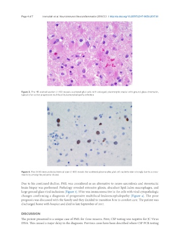
Figure 4. This SV40 immunohistochemical stain (×40) reveals the scattered pleomorphic glial cell nuclei to stain strongly due to a cross-
reactivity among the polyoma viruses
Due to his continued decline, PML was considered as an alternative to neuro-sarcoidosis and stereotactic
brain biopsy was performed. Pathology revealed extensive gliosis, abundant lipid-laden macrophages, and
large ground glass viral inclusions [Figure 3]. SV40 was immunoreactive in the cells with viral cytopathologic
changes confirming a diagnosis of progressive multifocal leukoencephalopathy [Figure 4]. The poor
prognosis was discussed with the family and they decided to transition him to comfort care. The patient was
discharged home with hospice and died in late September of 2017.
DISCUSSION
The patient presented is a unique case of PML for three reasons. First, CSF testing was negative for JC Virus
DNA. This caused a major delay in the diagnosis. Previous cases have been described where CSF PCR testing