
Page 29 - Read Online
P. 29
Inamullah et al. Neuroimmunol Neuroinflammation 2018;5:3 I http://dx.doi.org/10.20517/2347-8659.2017.61 Page 3 of 7
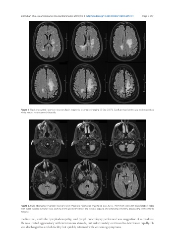
Figure 1. Fluid-attenuated inversion recovery brain magnetic resonance imaging (8 Sep 2017). Confluent periventricular and subcortical
white matter lesions seen bilaterally
Figure 2. Fluid-attenuated inversion recovery brain magnetic resonance imaging (8 Sep 2017). Prominent Wallerian degeneration noted
with lesion located in motor tract starting in the posterior limb of the internal capsule and extending inferiorly, decussating in the inferior
medulla
mediastinal, and hilar lymphadenopathy, and lymph node biopsy performed was suggestive of sarcoidosis.
He was treated aggressively with intravenous steroids, but unfortunately continued to deteriorate rapidly. He
was discharged to a rehab facility but quickly returned with worsening symptoms.