
Page 159 - Read Online
P. 159
Kafle et al. Neuroimmunol Neuroinflammation 2018;5:24 I http://dx.doi.org/10.20517/2347-8659.2018.10 Page 5 of 8
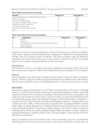
Table 4. Surgical procedures in the present study
Procedure Frequency (n) Percentage (%)
Burrhole and aspiration with modified radical mastoidectomy 23 45.08
Burrhole and aspiration 14 27.44
Burrhole and multiple aspiration 6 11.76
Craniotomy and subdural empyema drainage 3 5.88
Craniotomy and abscess wall excision 2 3.92
Continuous abscess drainage 2 3.92
Craniotomy and epidural abscess drainage 1 1.96
Total 51 100
Table 5. Complications observed in the study population
No. Complications Frequency (n) Percentage (%)
1 Mortality 2 3.92
2 Pyoventricle 1 1.96
3 Post modified radical mastoidectomy facial nerve palsy (grade II-V) 4 7.84
4 Pseudomeningocele 1 1.96
5 Surgical site infection 1 1.96
staphylococcus aureus. All cases were treated with 2 weeks of intravenous and 4 weeks of oral antibiotics
(Cefpodoxime 5 mg/kg 12 hourly and Metronidazole 25 mg/kg/day in 3 divided doses) including anaerobic
coverage. Some cases required 8 weeks of antibiotics. Oral versus intravenous route was determined
depending on the status of the abscess cavity on repeat head CT. Culture and sensitivity for anaerobic
organisms were not done in the present study due to resource constrains.
Complications
The mortality was 3.92% (n = 2) during the study. Major complications observed are listed in Table 5. The
most common minor complication noted was thrombophlebitis likely due to prolonged use of IV antibiotics.
Outcome
All surviving patients were followed up in outpatient clinic for at least 3 months. All of them had Glasgow
outcome scale of 5/5. Surgical site infection and pseudomeningocele was resolved at the 6 week follow up
visit. Facial palsy resolved in 3 cases, with residual palsy present in 1 case at follow-up cessation (3 months).
DISCUSSION
Brain abscess comprises approximately 8% of all space occupying lesions in the brain in developing
countries . Abscess is the second most common type of intracranial complication of otogenic origin, with
[1,2]
temporal lobe being the most common site of pathology. Clinical presentation varies among patients. The
classic triad of fever, headache and focal deficit is rarely seen. Features of raised intracranial pressure with
or without localizing signs require early radiological imaging to avoid inadvertent delay in management .
[4]
Contrast enhanced CT scan of the head is the mainstay of diagnostic modalities , providing rapid means
[5]
of detecting the lesion. MRI, combined with diffusion-weighted (DWI) and apparent-diffusion coefficient
(ADC) images, is a valuable diagnostic tool in differentiating brain abscess from primary, cystic, or necrotic
tumors with positive predictive value of 98% and negative predictive value of 92% . Cultures of blood and
[6]
cerebrospinal fluid identify the causative pathogen in approximately one quarter of patients. Cultures of
cerebrospinal fluid may be valuable in patients with coexisting meningitis. Lumbar puncture can lead to
herniation in such situations .
[7]
There is no pragmatic rule for the treatment of brain abscess. Treatment of each case is individualized
depending up on the location, size, and stage of abscess. The mainstay of treatment is prompt action and