
Page 158 - Read Online
P. 158
Page 4 of 8 Kafle et al. Neuroimmunol Neuroinflammation 2018;5:24 I http://dx.doi.org/10.20517/2347-8659.2018.10
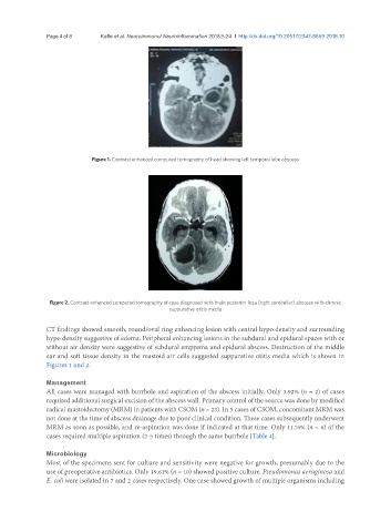
Figure 1. Contrast enhanced computed tomography of head showing left temporal lobe abscess
Figure 2. Contrast enhanced computed tomography of case diagnosed with brain posterior fosa (right cerebellar) abscess with chronic
suppurative otitis media
CT findings showed smooth, round/oval ring enhancing lesion with central hypo density and surrounding
hypo density suggestive of edema. Peripheral enhancing lesions in the subdural and epidural spaces with or
without air density were suggestive of subdural empyema and epidural abscess. Destruction of the middle
ear and soft tissue density in the mastoid air cells suggested suppurative otitis media which is shown in
Figures 1 and 2.
Management
All cases were managed with burrhole and aspiration of the abscess initially. Only 3.92% (n = 2) of cases
required additional surgical excision of the abscess wall. Primary control of the source was done by modified
radical mastoidectomy (MRM) in patients with CSOM (n = 23). In 5 cases of CSOM, concomitant MRM was
not done at the time of abscess drainage due to poor clinical condition. These cases subsequently underwent
MRM as soon as possible, and re-aspiration was done if indicated at that time. Only 11.76% (n = 6) of the
cases required multiple aspiration (2-5 times) through the same burrhole [Table 4].
Microbiology
Most of the specimens sent for culture and sensitivity were negative for growth, presumably due to the
use of preoperative antibiotics. Only 19.61% (n = 10) showed positive culture. Pseudomonas aeruginosa and
E. coli were isolated in 7 and 2 cases respectively. One case showed growth of multiple organisms including