
Page 26 - Read Online
P. 26
Oommen et al. Cavernous sinus cavernous hemangioma
suggestive of cavernous hemangioma [Figure 2]. identified two types of dural cavernous angiomas.
Follow-up (1) One present in the dura of the middle cranial fossa
The postoperative MRI brain with contrast did not usually in the vicinity of the cavernous sinus.
show any residual hemangioma [Figure 3]. The
patient was asymptomatic without any additional (2) The second type consists of dural-based lesions
neurologic deficit. such as the convexity, cerebral and cerebellar falx,
the tentorium, posterior fossa, and the floor of the
DISCUSSION anterior fossa. [4]
Cavernous angiomas are vascular malformations. The separation of the two types is important due to
They are composed of enlarged sinusoidal vessels the differences in the patient population affected and
arranged in clusters, enclosed by a thin endothelial wall the more aggressive clinical course of dural-based
without interposed tissue within. They lack an elastic cavernous angiomas in the middle cranial fossa.
lamina, smooth muscles and are sometimes ossified
or calcified. The term “cavernous angioma” has been The malformations of the first group are more clinically
used interchangeably with “cavernous hemangioma,” aggressive because of their localization and vascular
“cavernous malformation,” or “cavernoma”. These supply. In these cases, both preoperative radiation and
lesions are vascular abnormalities rather than embolization are recommended because they reduce
neoplastic processes. [1] intraoperative bleeding risk.
Cavernomas most commonly originates from the brain On the other hand, in the cases of the second group,
parenchyma. However, they also arise intraspinally, neither radiation nor embolization is necessary to
or from the dura. Extra-axial dural-based cavernous successfully remove cavernous hemangiomas outside
[2]
malformations are extremely rare when compared to the middle cranial fossa, since their vascular supply can
their intra-axial counterparts. In 1994, Lewis et al. be easily controlled through the surgical exposure. [5]
[3]
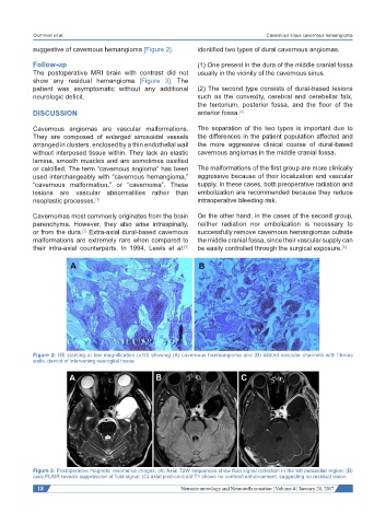
Figure 2: HE staining at low magnification (×10) showing (A) cavernous haemangioma and (B) dilated vascular channels with fibrous
walls, devoid of intervening neuroglial tissue
Figure 3: Postoperative magnetic resonance images. (A) Axial T2W sequences show fluid signal collection in the left parasellar region; (B)
axial FLAIR reveals suppression of fluid signal; (C) axial post-contrast T1 shows no contrast enhancement, suggesting no residual lesion
18 Neuroimmunology and Neuroinflammation ¦ Volume 4 ¦ January 20, 2017