
Page 25 - Read Online
P. 25
Oommen et al. Cavernous sinus cavernous hemangioma
Imaging was done using ultrasonic aspirator, suction and
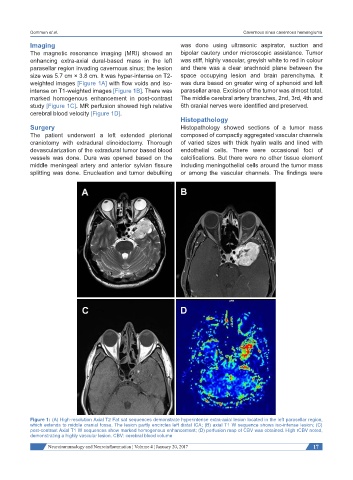
The magnetic resonance imaging (MRI) showed an bipolar cautery under microscopic assistance. Tumor
enhancing extra-axial dural-based mass in the left was stiff, highly vascular, greyish white to red in colour
parasellar region invading cavernous sinus; the lesion and there was a clear arachnoid plane between the
size was 5.7 cm × 3.8 cm. It was hyper-intense on T2- space occupying lesion and brain parenchyma. It
weighted images [Figure 1A] with flow voids and iso- was dura based on greater wing of sphenoid and left
intense on T1-weighted images [Figure 1B]. There was parasellar area. Excision of the tumor was almost total.
marked homogenous enhancement in post-contrast The middle cerebral artery branches, 2nd, 3rd, 4th and
study [Figure 1C]. MR perfusion showed high relative 6th cranial nerves were identified and preserved.
cerebral blood velocity [Figure 1D].
Histopathology
Surgery Histopathology showed sections of a tumor mass
The patient underwent a left extended pterional composed of compactly aggregated vascular channels
craniotomy with extradural clinoidectomy. Thorough of varied sizes with thick hyalin walls and lined with
devascularization of the extradural tumor based blood endothelial cells. There were occasional foci of
vessels was done. Dura was opened based on the calcifications. But there were no other tissue element
middle meningeal artery and anterior sylvian fissure including meningothelial cells around the tumor mass
splitting was done. Enucleation and tumor debulking or among the vascular channels. The findings were
Figure 1: (A) High-resolution Axial T2 Fat sat sequences demonstrate hyperintense extra-axial lesion located in the left parasellar region,
which extends to middle cranial fossa. The lesion partly encircles left distal ICA; (B) axial T1 W sequence shows iso-intense lesion; (C)
post-contrast Axial T1 W sequences show marked homogenous enhancement; (D) perfusion map of CBV was obtained. High rCBV noted,
demonstrating a highly vascular lesion. CBV: cerebral blood volume
Neuroimmunology and Neuroinflammation ¦ Volume 4 ¦ January 20, 2017 17