
Page 214 - Read Online
P. 214
Infante et al. PML after rituximab
dose administered two years before PML onset)
without signs of hematological abnormalities; and (3)
rapid neurological deterioration (day-by-day worsening
with “sudden” onset of the symptoms) made PML
diagnosis particularly tricky. Despite single atypical
characteristics have already been reported in the
literature, association of such findings have never
been reported so far.
As shown by MRI and CT scan, absence of a distinctive
vascular territory lesion distribution, despite the acute
clinical course and the posterior fossa localization
could have been an early clue to a correct diagnosis.
In this case, diagnosis of PML has been delayed
because of the clinical presentation (acute onset of
cerebellar symptoms), initial lack of anamnestic data
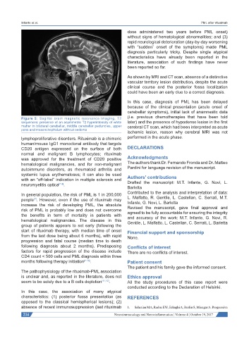
Figure 5: Sagittal brain magnetic resonance imaging, T2 (i.e. previous chemotherapies that have been told
sequences: presence of an asymmetric T2 hyperintensity of white later) and the presence of hypodense lesion in the first
matter in bilateral cerebellar, middle cerebellar peduncles, upper cerebral CT scan, which had been interpreted as acute
pons and mesenchephalum without oedema
ischemic lesion, reason why cerebral MRI was not
lymphoproliferative disorders. Rituximab is a chimeric performed in the acute phase.
human/mouse IgG1 monoclonal antibody that targets
CD20 antigen expressed on the surface of both DECLARATIONS
normal and malignant B lymphocytes; rituximab
was approved for the treatment of CD20 positive Acknowledgments
hematological malignancies, and for non-malignant The authors thank Dr. Fernando Fronda and Dr. Matteo
autoimmune disorders, as rheumatoid arthritis and Pardini for language revision of the manuscript.
systemic lupus erythematosus; it can also be used
with an “off-label” indication in multiple sclerosis and Authors’ contributions
Drafted the manuscript: M.T. Infante, G. Novi, L.
neuromyelitis optica [7-9] .
Barletta
In general population, the risk of PML is 1 in 200,000 Contributed to the analysis and interpretation of data:
people . However, even if the use of rituximab may L. Malfatto, R. Gentile, L. Castellan, C. Serrati, M.T.
[7]
increase the risk of developing PML, the absolute Infante, G. Novi, L. Barletta
risk of PML is probably low and does not overcome Revised the manuscript, gave final approval and
the benefits in term of mortality in patients with agreed to be fully accountable for ensuring the integrity
and accuracy of the work: M.T. Infante, G. Novi, R.
hematological malignancies. The disease in this Gentile, L. Malfatto, L. Castellan, C. Serrati, L. Barletta
group of patients appears to set early (following the
start of rituximab therapy, with median time of onset Financial support and sponsorship
from the last dose being about 6 months), with rapid None.
progression and fatal course (median time to death
following diagnosis about 2 months). Predisposing Conflicts of interest
factors for rapid progression of the disease include There are no conflicts of interest.
CD4 count < 500 cells and PML diagnosis within three
months following therapy initiation [7-10] . Patient consent
The patient and his family gave the informed consent.
The pathophysiology of the rituximab-PML association
is unclear and, as reported in the literature, does not Ethics approval
seem to be solely due to a B cells depletion [11,12] . All the study procedures of this case report were
conducted according to the Declaration of Helsinki.
In this case, the association of many atypical
characteristics: (1) posterior fossa presentation (as REFERENCES
opposed to the classical hemispherical lesions); (2)
absence of recent immunosuppression (last rituximab 1. Sahraian MA, Radue EW, Eshaghi A, Besliu S, Minagar A. Progressive
214 Neuroimmunology and Neuroinflammation ¦ Volume 4 ¦ October 19, 2017