
Page 197 - Read Online
P. 197
Markoula et al. Restricted diffusiuon in mulltpile sclerosis lesions
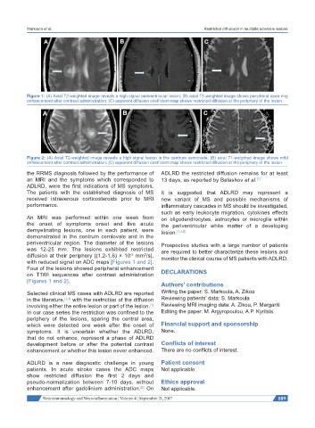
Figure 1: (A) Axial T2-weighted image reveals a high signal periventricular lesion; (B) axial T1-weighted image shows peripheral open ring
enhancement after contrast administration; (C) apparent diffusion coefficient map shows restricted diffusion at the periphery of the lesion
Figure 2: (A) Axial T2-weighted image reveals a high signal lesion in the centrum semiovale; (B) axial T1-weighted image shows mild
enhancement after contrast administration; (C) apparent diffusion coefficient map shows restricted diffusion at the periphery of the lesion
the RRMS diagnosis followed by the performance of ADLRD the restricted diffusion remains for at least
an MRI and the symptoms which corresponded to 13 days, as reported by Balashov et al. [1]
ADLRD, were the first indications of MS symptoms.
The patients with the established diagnosis of MS It is suggested that ADLRD may represent a
received intravenous corticosteroids prior to MRI new variant of MS and possible mechanisms of
performance. inflammatory cascades in MS should be investigated,
such as early leukocyte migration, cytokines effects
An MRI was performed within one week from on oligodendrocytes, astrocytes or microglia within
the onset of symptoms onset and five acute the periventricular white matter of a developing
demyelinating lesions, one in each patient, were lesion. [1,4,5]
demonstrated in the centrum cemiovale and in the
periventricular region. The diameter of the lesions Prospective studies with a large number of patients
was 12-25 mm. The lesions exhibited restricted are required to better characterize these lesions and
diffusion at their periphery [(1.2-1.6) × 10 mm /s], monitor the clinical course of MS patients with ADLRD.
2
-3
with reduced signal on ADC maps [Figures 1 and 2].
Four of the lesions showed peripheral enhancement
on T1WI sequences after contrast administration DECLARATIONS
[Figures 1 and 2].
Authors’ contributions
Selected clinical MS cases with ADLRD are reported Writing the paper: S. Markoula, A. Zikou
in the literature, [3,4] with the restriction of the diffusion Reviewing patients’ data: S. Markoula
involving either the entire lesion or part of the lesion. Reviewing MRI imaging data: A. Zikou, P. Margariti
[1]
In our case series the restriction was confined to the Editing the paper: M. Argyropoulou, A.P. Kyritsis
periphery of the lesions, sparing the central area,
which were detected one week after the onset of Financial support and sponsorship
symptoms. It is uncertain whether the ADLRD, None.
that do not enhance, represent a phase of ADLRD
development before or after the potential contrast Conflicts of interest
enhancement or whether this lesion never enhanced. There are no conflicts of interest.
ADLRD is a new diagnostic challenge in young Patient consent
patients. In acute stroke cases the ADC maps Not applicable.
show restricted diffusion the first 2 days and
pseudo-normalization between 7-10 days, without Ethics approval
enhancement after gadolinium administration. On Not applicable.
[2]
Neuroimmunology and Neuroinflammation ¦ Volume 4 ¦ September 21, 2017 189