
Page 11 - Read Online
P. 11
Chu et al. Meningeal carcinomatosis: a retrospective analysis
cases showed abnormal cells; while in 8.2% (6/73) the increased CA125 was 32% (8/25). 32% (8/25) of
cases abnormal cells were found through a second the patients had increased CA199. In 28% (7/25) of
time lumbar puncture; only in one case tumor the patients NSE increase was observed. The rate
cells were found in the third lumbar puncture. Cell of the increased CA153 was 24% (6/25). There were
morphology analysis revelaed that 14 cases showed 16% (4/25) of the patients who had increased CA724.
adenocarcinoma cells. The remaining cases were Four percent (1/25) of the patients’ alpha-fetoprotein
characterized by an increase in cell size, irregular increased. The rate of elevated blood sedimentation
shape, cell body stain and darker cytoplasm. Cell was 66.7% (10/15).
membranes were incomplete and with protrusions.
Nuclear cytoplasm ratio increased, and nucleus were Imaging findings in MC patients
centered or showed deviation, occasionally they were Twenty-four cases had head computed tomography
double-nucleated. In some cells, cytoplasmic vacuoles examinations. Among these, one of them showed that
were observed near the membrane. Some mitotic meninges thickened significantly. One case showed
cells could also be observed. The cytology results for the expansion of the ventricles and hydrocephalus,
different cancers are shown in Figure 2. Only 3 cases the others had no significant abnormalities. Fifty-
of the CSF tumor markers were checked, and all the three cases had head MRI scan examinations and 17
results were abnormal. Patients with elevated IgG (32.1%) of them were abnormal. A total of 12 (35.3%)
accounted for 72.7% (16/22). cases had meningeal reinforcement in the 34 cases of
CSF routine biochemical examination was found enhanced scan.
abnormal in 95.3% (61/64) patients. High protein Primary tumors
accounted for 74% (46/62) (normal range 0.15-0.45 g/L),
reduced glucose was 45% (27/60) (normal range 2.3- The most frequent primary tumor in our study was
4.1 mmol/L), reduced chlorine accounted for 38% lung cancer (35/77, 58.3%), followed by gastric cancer
(23/60) (normal range 119-129 mmol/L), elevated white (10/77, 16.7%), breast cancer (6/77, 10%), melanoma
blood cell count was 59.7% (37/62) (normal range 0-8 (3/77, 5%) and non-hodgkin’s lymphoma (2/77, 3.3%).
6
× 10 /L), which was given priority to mononuclear cells. In addition, one patient presented with primary lesion
in ovarian and one presented with colon cancer. In
Tumor marker changes in MC patients one case the primary tumor was nasopharyngeal
The rate of abnormal serum tumor markers was carcinoma, and in another it was acute lymphocytic
84%. Sixty-eight percent (17/25) of the patients had leukemia. Seventeen (22.1%) had no primary tumor.
increased carcinoembryonic antigen. In 44% (11/25) The interval from diagnosis of primary tumor to the
of the patients CYFRA21-1 increased. The rate of onset of central nervous system (CNS) symptoms was
also analyzed: 45 cases (58.4%) initially presented
with CNS symptoms without history of tumor; 26%
(20/77) patients developed CNS symptoms when the
primary tumor had been diagnosed for no more than
one year; 6.5% (5/77) patients experienced CNS
symptoms at one to two years after the diagnosis of
primary tumor; 9.1% (7/77) patients did not experience
the CNS symptoms until the primary tumor has been
diagnosed for more than two years.
Treatment and survival
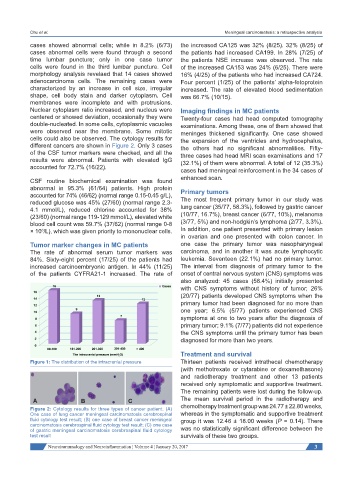
Figure 1: The distribution of the intracranial pressure Thirteen patients received intrathecal chemotherapy
(with methotrexate or cytarabine or dexamethasone)
and radiotherapy treatment and other 13 patients
received only symptomatic and supportive treatment.
The remaining patients were lost during the follow-up.
The mean survival period in the radiotherapy and
chemotherapy treatment group was 24.77 ± 22.80 weeks,
Figure 2: Cytology results for three types of cancer patient. (A)
One case of lung cancer meningeal carcinomatosis cerebrospinal whereas in the symptomatic and supportive treatment
fluid cytology test result; (B) one case of breast cancer meningeal group it was 12.46 ± 18.00 weeks (P = 0.14). There
carcinomatosis cerebrospinal fluid cytology test result; (C) one case
of gastric meningeal carcinomatosis cerebrospinal fluid cytology was no statistically significant difference between the
test result survivals of these two groups.
Neuroimmunology and Neuroinflammation ¦ Volume 4 ¦ January 20, 2017 3