
Page 159 - Read Online
P. 159
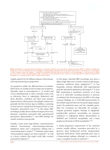
Figure 2. Flowchart for approaching the recognition of encephalitis related to neuronal surface antibody syndrome (NSAS) in children [modified from
                                     [8]
                    [1]
            Zuliani et al.  2012 and Suleiman et al. 2013]. NSAbs: neuronal surface antibodies; CSF: cerebrospinal fluid; CXCL13: chemokine (C-X-C motif) ligand
            13; MRI: magnetic resonance imaging; *: in NMDA-R encephalitis; GAD-Abs: antibodies to glutamic acid decarboxylase; *: GAD positivity is defined
                        [8]
            as > 1,000 u/mL.  OCB: oligoclonal bands; IvMP: intravenous methylprednisolone; IvIg: intravenous immunoglobulin; PE: plasma exchange; RTX:
            Rituximab; Cyc: cyclophosphamide; MMF: mycophenolate mophetil.
            roughly specific for the different phases of the disease,   In this phase, interictal EEG recordings may show a
            allowing monitoring its progression.              diffuse high delta waves activity with focal discharges,
                                                              refractory multifocal status epilepticus [38,39]  or less
            As reported in adults, the different forms of pediatric   frequently, extreme delta-brush with superimposed
            NSAS share an overlap of clinical signs and symptoms.   periodic lateralized epileptiform discharges.  A close
                                                                                                    [39]
            Typically, onset is acute/subacute (< 12 weeks) and   EEG monitoring is mandatory, inclusive of at least
            occurs simultaneously or after a stressful event, such   one 24 h video-EEG recording (duration to increase
            as  infections,  fever  or  cephalalgia.  Behavioral  and   according to the clinical picture). On MRI, in this stage
            sleep disorders, confusion and short term memory   common findings are a T2/FLAIR hyperintensity in
            impairment are often present, but epileptic seizures are   the medial temporal lobes involving the hippocampus,
            generally the first obvious sign in children, consisting
            in focal seizures arising from bi-temporal lobes or, less   and/or the prefrontal areas and the cingulate gyrus.
            frequently, generalized ones. [8-10]  In this phase EEG   Only few exceptions are detectable, for example a
            recordings usually shows a diffuse background activity   frequent persistent  negative findings in  dipeptidyl-
            slowness, sometimes associated with superimposed   peptidase-like protein-6 encephalitis, an extensive
            paroxysmal abnormalities, [9,39]  and MRI findings are   multifocal or widespread diffuse abnormalities in
            usually normal or non specific.                   NMDAR and GABAA-R encephalitis, and a basal
                                                              ganglia involvement in D2-R encephalitis. [42,43]
            Usually, a more acute stage follows, characterized by
            reduced consciousness, refractory/super-refractory   A slowly progressive improvement of the interictal
            epilepticus status and a progressive sinking into a   activity usually follows, sometimes preceded by a
            coma lasting days to weeks. [40,41]  Ventilator and/or other   previous  sleep  background  activity  reorganization
                                                  [13]
            vital parameters support are often required,  as well   (personal observation), which generally gives way to
            as a heavy sedation that might easily hide eventual   a  drug-resistant epilepsy  although, in  a  minority  of
            associated symptoms. [28]                         cases, a complete recovery is achieved. [44]
             150                                                  Neuroimmunol Neuroinflammation | Volume 3 | July 8, 2016