
Page 18 - Read Online
P. 18
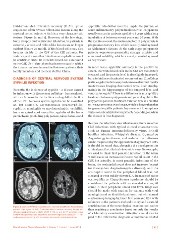
fluid-attenuated inversion recovery  (FLAIR) pulse   syphilitic retrobulbar neuritis), syphilitic gumma, or
          sequences, often reveals ribbon-like lesions along the   acute inflammatory polyradiculoneuritis. Polyparesis
          cerebral cortex lesions, which is a very characteristic   usually occurs in patients aged 35–45 years with a long
          feature [Figure 2a and b]. However, at the late stage,   incubation of between several years and 20 years. With
          brain atrophy and ventricular dilatation in patients is   the insidious onset, the main symptom of polyparesis is
          extremely severe, and ribbon-like lesions are no longer   progressive memory loss, which is easily misdiagnosed
          evident [Figure 2c and d]. White blood cells may also   as Alzheimer’s disease. At the early stage, polyparesis
          become visible in the CSF of the CJD patients. For   patients experience personality changes, anxiety, and
          instance, a virus or other infectious encephalitis cannot   emotional volatility, which can easily be misdiagnosed
          be confirmed until 10–30 white blood cells are found   as depression.
          in the CSF. Until date, there has been no case in which
          the disease has been transmitted between patients, their   In most cases, syphilitic antibody is the positive in
          family members and medical staff in China.          serum, the white blood cells in the CSF are moderately
                                                              elevated, and the protein level is also slightly increased,
          DIAGNOSIS OF CENTRAL NERVOUS SYSTEM                 but a toluidine red unheated serum test and T. pallidum
          SYPHILIS INFECTION                                  particle agglutination assay test can reveal normal results
                                                              in a few cases. Imaging observations reveal brain atrophy,
          Recently, the incidence of syphilis – a disease caused   mainly in the hippocampus of the temporal lobe, and
                                                                              [7]
          by infection with Treponema pallidum – has escalated,   ventriculomegaly.  There is a difference in antisyphilitic
          with an increase in the incidence of syphilis infection   treatment between polyparesis and general syphilis. For
          of the CNS. Nervous system syphilis can be classified   polyparesis patients, treatment duration time is 6 months
          as, for example, asymptomatic neurosyphilis,        to 1 year, sometimes even longer, which is longer than that
          syphilitic meningitis or myelomeningitis, syphilitic   for general syphilis patients. Improvement in symptoms
          brain or spinal cord vasculitis, syphilis of the brain   varies considerably between patients depending on when
          parenchyma (including polyparesis, tabes dorsalis and   the disease is first diagnosed.
                                                              Besides the infections described above, there are other
                                                              CNS infections with typical clinical characteristics,
                                                              such as human immunodeficiency virus, Brinell
                                                              bacillus infection, Whipple’s disease, Guangzhou
                                                              Angiostrongylus disease, and malaria. Such diseases
                                                              can be diagnosed by the application of appropriate tests.
                                                              It should be noted that, alongside the development in
                                                              clinical practice, clinical viewpoints vary. For example,
                                                              we used to think that parasitic infection in the brain
                                                              would cause an increase in the eosinophil count in the
           a                        b                         CSF, but actually, in most parasitic infections of the
                                                              brain, the eosinophil count does not increase (except
                                                              for Guangzhou Angiostrongylus disease), and the
                                                              eosinophil count in the peripheral blood was not
                                                              elevated or even mildly elevated. A diagnosis of either
                                                              eosinophilia or Churg–Strauss syndrome should be
                                                              considered for patients with an elevated eosinophil
                                                              count in their peripheral blood and fever. Diagnoses
                                                              should be made with caution for patients with viral
                                                              meningitis and no identified pathogen, but with normal
                                                              electroencephalography, brain MRI scan and CSF, with
           c                      d                           reference to the patient’s medical history and a careful
          Figure 2: (a and b) MRI representations of a sporadic Creutzfeldt–Jakob disease   consideration of the neurological examination, rather
          patient:  high  signal  on  both  fluid-attenuated  inversion  recovery  image  and   than reaching a conclusion based on only the results
          diffusion-weighted imaging (DWI) (2009–6–16). (c and d) T2–weighted image   of a laboratory examination. Attention should also be
          and DWI image of the same patient in a persistent vegetative state (2011–7–13):
          serious encephalatrophy with an obvious increase in ventricular volume  paid to the differential diagnosis of immune-mediated


          Neuroimmunol Neuroinflammation | Volume 1 | Issue 1 | June 2014                                   11