
Page 111 - Read Online
P. 111
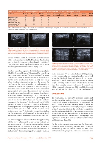
Table 2:  An overview of the existing nerve ultrasound studies on MMN and their pathological findings
           Authors          Patients   Controls   Median   Ulnar   Brachial plexus   Sciatic   Femoral   Fibular   Tibial
                              (n)       (n)     nerve   nerve   or cervical roots  nerve  nerve   nerve   nerve
           Beekman et al. [6]  21       20        x       x           x           ‑        ‑        ‑       ‑
           Padua et al. [7]    2        63        x       x           ‑           ‑        ‑        x       ‑
           Kerasnoudis [8]     2        30        x       x           ‑           ‑        ‑        x       ‑
           Kerasnoudis et al. [9]  1     ‑        x       N          N            ‑        ‑        ‑       ‑
           Kerasnoudis et al. [10]  12  80        x       x          N            ‑        ‑       N        x
           Zaidman et al. [14]  17       ‑        x       x           ‑           ‑        ‑        ‑       ‑
           MMN: Multifocal motor neuropathy; x: The concrete nerve was reported with pathological findings; N: The concrete nerve was reported with normal findings; -: The
           concrete nerve was not studied at all; n: Absolute number


           a                       b                           a                        b
           Figure 1: Axial scan of the brachial plexus in a multifocal motor neuropathy
           (MMN) patient in supraclavicular (a) and interscalene space (b) in a MMN patient.
           In this case both the cross sectional area and the intraplexus cross sectional
           area variability of the brachial plexus are within the reference values of our lab
                                                         [13]
           several proximal and distal sites in the anatomic course
           of the peripheral nerves in MMN patients. This finding
           may reflect the immune-mediated patchy multifocal   c                        d
           demyelination occurring along the motor nerve fibers   Figure 2: Axial scan of the ulnar nerve in a multifocal motor neuropathy (MMN)
                                                              patient in Guyon’s canal (a), forearm (b), elbow (c) and upper‑arm (d) showing
           in this type of immune-mediated injury. [10,11]    a pathological cross sectional area enlargement at all anatomic sites, when
                                                                                           [13]
                                                              compared to the reference values of our lab.  This finding may reflect the
                                                              immune‑mediated patchy multifocal demyelination occurring in MMN
           Another important aspect in the field of sonography in
           MMN is the possible use of this method for identifying   in the literature. [16,17]  In a later study on MMN patients,
           nerve conduction blocks. The localization of the nerve   neither sonography nor electrophysiology correlated
           conduction block is often difficult to be identified   with the Medical  Research Council  sum  score,
           in the nerve conduction studies  (NCS), especially   Rasch-built Overall Disability Scale score or Rasch-built
           when dealing with proximal parts of the nerves. By   fatigue severity scale. [10]  These studies have shown
           overlooking the electrophysiological hallmark of the   that the already known ultrasound biomarkers
           disease, delay in the diagnosis and therefore delayed   (CSA, echogenity, intranerve CSA variability) are not
           treatment can occur.  Beekman et al.  documented   able to highlight the effectivity of immune-therapy. [11]
                                             [6]
                             [2]
           pathological ultrasound findings not only at sites
           with  electrophysiological  impairment,  but  also  at   CONCLUSION
           sites with normal functioning in NCS. An absolute
           correlation between site of nerve hypertrophy and   To summarize, the currently available ultrasound
           site of conduction block has been reported only in   studies show that mainly a focal type of asymmetrical
                                 [8]
           one case in the literature.  Another study on 12 MMN   peripheral  nerve  enlargement  is  expected  in
           patients showed a significant correlation between   MMN. Nerve ultrasound findings seem to show no
           sonographic and electrophysiological findings only   significant correlation to electrophysiological findings
           between  the  CMAP  and  CSA  of  the  median  nerve   at most anatomical sites. In addition, prospective
           at the upper arm. [10]  Systematic prospective studies   studies on the applicability of ultrasound as screening
           on the sensitivity of ultrasound in detecting focal   method of immune-therapy fail in the literature, while
           immune-mediated nerve lesions fail in the literature.  various retrospective studies failed to highlight any
                                                              significant correlation between ultrasound findings
           An interesting point of future study is the applicability   and functional disability.
           of the nerve ultrasound as screening method for
           immune-therapy in dysimmune neuropathies. Nerve    As the main uncertainties regarding the diagnostic
           ultrasound and NCS failed to highlight functional   criteria of MMN are steadily resolved, new challenges
           disability in post-Guillain-Barré syndrome and chronic   continuously arise on how to acquire the best static
           inflammatory demyelinating polyneuropathy patients   and dynamic imaging of the relevant nerve structures


          Neuroimmunol Neuroinflammation | Volume 1 | Issue 3 | December 2014                               105