
Page 84 - Read Online
P. 84
Wiens et al. Mini-invasive Surg 2021;5:8 I http://dx.doi.org/10.20517/2574-1225.2020.105 Page 5 of 7
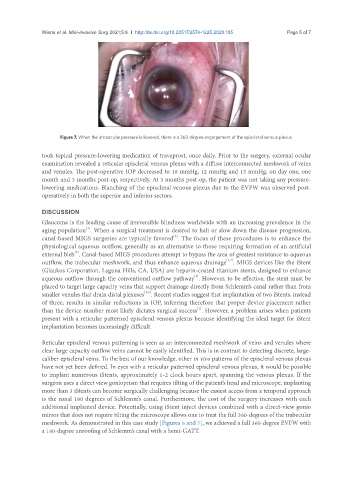
Figure 7. When the intraocular pressure is lowered, there is a 360-degree engorgement of the episcleral venous plexus.
took topical pressure-lowering medication of travaprost, once daily. Prior to the surgery, external ocular
examination revealed a reticular episcleral venous plexus with a diffuse interconnected meshwork of veins
and venules. The post-operative IOP decreased to 18 mmHg, 12 mmHg and 15 mmHg, on day one, one
month and 3 months post-op, respectively. At 3 months post-op, the patient was not taking any pressure-
lowering medications. Blanching of the episcleral venous plexus due to the EVFW was observed post-
operatively in both the superior and inferior sectors.
DISCUSSION
Glaucoma is the leading cause of irreversible blindness worldwide with an increasing prevalence in the
[1]
aging population . When a surgical treatment is desired to halt or slow down the disease progression,
canal-based MIGS surgeries are typically favored . The focus of these procedures is to enhance the
[6]
physiological aqueous outflow, generally as an alternative to those requiring formation of an artificial
external bleb . Canal-based MIGS procedures attempt to bypass the area of greatest resistance to aqueous
[6]
[1,7]
outflow, the trabecular meshwork, and thus enhance aqueous drainage . MIGS devices like the iStent
(Glaukos Corporation, Laguna Hills, CA, USA) are heparin-coated titanium stents, designed to enhance
[2]
aqueous outflow through the conventional outflow pathway . However, to be effective, the stent must be
placed to target large capacity veins that support drainage directly from Schlemm’s canal rather than from
[2,8]
smaller venules that drain distal plexuses . Recent studies suggest that implantation of two iStents, instead
of three, results in similar reductions in IOP, inferring therefore that proper device placement rather
[1]
than the device number most likely dictates surgical success . However, a problem arises when patients
present with a reticular patterned episcleral venous plexus because identifying the ideal target for iStent
implantation becomes increasingly difficult.
Reticular episcleral venous patterning is seen as an interconnected meshwork of veins and venules where
clear large capacity outflow veins cannot be easily identified. This is in contrast to detecting discrete, large-
caliber episcleral veins. To the best of our knowledge, other in vivo patterns of the episcleral venous plexus
have not yet been defined. In eyes with a reticular patterned episcleral venous plexus, it would be possible
to implant numerous iStents, approximately 1-2 clock hours apart, spanning the venous plexus. If the
surgeon uses a direct view gonioprism that requires tilting of the patient’s head and microscope, implanting
more than 3 iStents can become surgically challenging because the easiest access from a temporal approach
is the nasal 180 degrees of Schlemm’s canal. Furthermore, the cost of the surgery increases with each
additional implanted device. Potentially, using iStent inject devices combined with a direct-view gonio
mirror that does not require tilting the microscope allows one to treat the full 360-degrees of the trabecular
meshwork. As demonstrated in this case study [Figures 6 and 7], we achieved a full 360-degree EVFW with
a 180-degree unroofing of Schlemm’s canal with a hemi-GATT.