
Page 82 - Read Online
P. 82
Wiens et al. Mini-invasive Surg 2021;5:8 I http://dx.doi.org/10.20517/2574-1225.2020.105 Page 3 of 7
Figure 1. Hemi-gonioscopy assisted transluminal trabeculotomy performed with ripcord technique in the inferior sector.
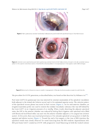
Figure 2. External ocular examination post-hemi-gonioscopy assisted transluminal trabeculotomy showed a reticular episcleral venous
pattern in the superior and inferior sectors. Note that the reticular pattern is more pronounced in the superior sector (left of image).
Figure 3. Reducing the intraocular pressure results in engorgement of the episcleral venous plexus superiorly and inferiorly.
[3,6]
The procedure for EVFW generation, as described below, was based on that described by Fellman et al. .
Post-hemi-GATT, the gonioscopy lens was removed for external examination of the episcleral vasculature
both adjacent to the treated site (inferior sector) and in the untreated superior sector. The reticular pattern
of the episcleral venous plexus was noted in both sectors [Figure 2]. In the mid-anterior chamber, an
irrigation/aspiration probe was used to remove the residual viscoelastic substance that was left behind by
the hemi-GATT at an irrigation pressure of 65 mmHg. When closely observing the adjacent episcleral
veins, the IOP in the anterior chamber was reduced by halting fluid irrigation until episcleral veins filled
with blood and focal blood reflux was seen into the anterior chamber adjacent to the surgical site (inferior
sector). At this point, there was maximal prominence of the reticular episcleral venous pattern in both the
superior and inferior sectors [Figure 3]. Toward the end of the surgery, at the time of BSS injection, the
episcleral vessels were closely observed for vessel blanching from the BSS washout. Hyperinfusion with
BSS post-hemi-GATT created an EVFW with progressive vessel blanching in both the treated inferior