
Page 83 - Read Online
P. 83
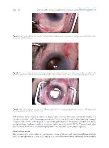
Page 4 of 7                                      Wiens et al. Mini-invasive Surg 2021;5:8  I  http://dx.doi.org/10.20517/2574-1225.2020.105


               Figure 4. Hyperinfusion of the anterior chamber with balanced salt solution causes an episcleral venous fluid wave and blanching of both
               the superior and inferior sectors.


               Figure 5. Reducing the intraocular pressure to near-physiologic conditions results in a prominent reticular episcleral venous pattern in the
               superior, untreated sector (left of image). Note that there is still some residual blanching in the inferior, treated sector (right of image).


               Figure 6. Hyperinflation of the anterior chamber with balanced salt solution to supraphysiologic conditions results in a 360-degree limbal
               blanching during the episcleral venous fluid wave.


               and untreated superior sectors [Figure 4]. Reducing IOP to near-physiologic conditions resulted in a
               prominent reticular episcleral venous pattern in the superior, untreated sector; residual blanching remained
               in the treated, inferior sector [Figure 5]. Repeated hyperinflation of the anterior chamber with BSS to
               supraphysiologic conditions results in 360-degree limbal blanching during EVFW [Figure 6], and when
               IOP is lowered, there was a 360-degree engorgement of the episcleral venous plexus [Figure 7].


               Results/Case study
               This procedure was performed on the right eye of a 58-year-old female with pigmentary glaucoma in both
               eyes. The pre-operative IOP was 20/21 mmHg as measured with Goldmann tonometry, and the subject