
Page 70 - Read Online
P. 70
Page 2 of 11 Elefante et al. Mini-invasive Surg 2021;5:7 I http://dx.doi.org/10.20517/2574-1225.2020.102
A B C
D E F
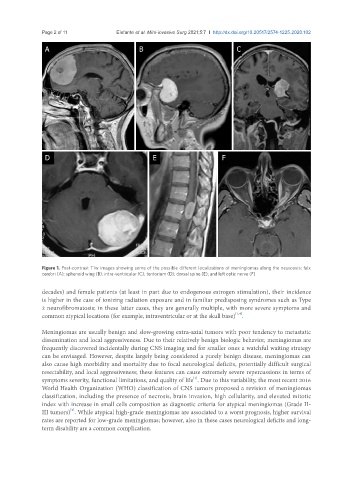
Figure 1. Post-contrast T1w images showing some of the possible different localizations of meningiomas along the neuroaxis: falx
cerebri (A); sphenoid wing (B); intra-ventricular (C); tentorium (D); dorsal spine (E); and left optic nerve (F)
decades) and female patients (at least in part due to endogenous estrogen stimulation), their incidence
is higher in the case of ionizing radiation exposure and in familiar predisposing syndromes such as Type
2 neurofibromatosis; in these latter cases, they are generally multiple, with more severe symptoms and
[1,4]
common atypical locations (for example, intraventricular or at the skull base) .
Meningiomas are usually benign and slow-growing extra-axial tumors with poor tendency to metastatic
dissemination and local aggressiveness. Due to their relatively benign biologic behavior, meningiomas are
frequently discovered incidentally during CNS imaging and for smaller ones a watchful waiting strategy
can be envisaged. However, despite largely being considered a purely benign disease, meningiomas can
also cause high morbidity and mortality due to focal neurological deficits, potentially difficult surgical
resectability, and local aggressiveness; these features can cause extremely severe repercussions in terms of
symptoms severity, functional limitations, and quality of life . Due to this variability, the most recent 2016
[1]
World Health Organization (WHO) classification of CNS tumors proposed a revision of meningiomas
classification, including the presence of necrosis, brain invasion, high cellularity, and elevated mitotic
index with increase in small cells composition as diagnostic criteria for atypical meningiomas (Grade II-
[5]
III tumors) . While atypical high-grade meningiomas are associated to a worst prognosis, higher survival
rates are reported for low-grade meningiomas; however, also in these cases neurological deficits and long-
term disability are a common complication.