
Page 73 - Read Online
P. 73
Elefante et al. Mini-invasive Surg 2021;5:7 I http://dx.doi.org/10.20517/2574-1225.2020.102 Page 5 of 11
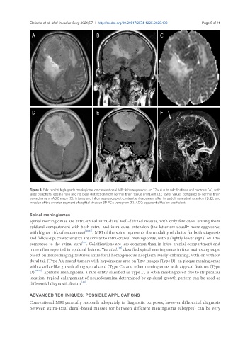
A B C
D E F
Figure 3. Falx cerebri high-grade meningioma on conventional MRI: inhomogeneous on T2w due to calcifications and necrosis (A), with
large peripheral edema halo and no clear distinction from normal brain tissue on FLAIR (B); lower values compared to normal brain
parenchyma on ADC maps (C); intense and inhomogeneous post-contrast enhancement after i.v. gadolinium administration (D, E); and
invasion of the anterior segment of sagittal sinus on 3D PCA venogram (F). ADC: apparent diffusion coefficient
Spinal meningiomas
Spinal meningiomas are extra-spinal intra-dural well-defined masses, with only few cases arising from
epidural compartment with both extra- and intra-dural extension (the latter are usually more aggressive,
with higher risk of recurrence) [26,27] . MRI of the spine represents the modality of choice for both diagnosis
and follow-up; characteristics are similar to intra-cranial meningiomas, with a slightly lower signal on T2w
[28]
compared to the spinal cord . Calcifications are less common than in intra-cranial compartment and
more often reported in epidural lesions. Yeo et al. classified spinal meningiomas in four main subgroups,
[29]
based on neuroimaging features: intradural homogeneous neoplasm avidly enhancing, with or without
dural tail (Type A); round tumors with hypointense area on T2w images (Type B); en plaque meningiomas
with a collar-like growth along spinal cord (Type C); and other meningiomas with atypical features (Type
D) [29,30] . Epidural meningioma, a rare entity classified as Type D, is often misdiagnosed due to its peculiar
location; typical enlargement of neuroforamina determined by epidural growth pattern can be used as
[31]
differential diagnostic feature .
ADVANCED TECHNIQUES: POSSIBLE APPLICATIONS
Conventional MRI generally responds adequately to diagnostic purposes, however differential diagnosis
between extra-axial dural-based masses (or between different meningioma subtypes) can be very