
Page 72 - Read Online
P. 72
Page 4 of 11 Elefante et al. Mini-invasive Surg 2021;5:7 I http://dx.doi.org/10.20517/2574-1225.2020.102
A B C
D E F
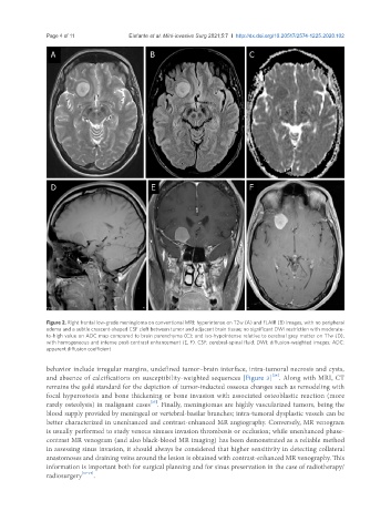
Figure 2. Right frontal low-grade meningioma on conventional MRI: hyperintense on T2w (A) and FLAIR (B) images, with no peripheral
edema and a subtle crescent-shaped CSF cleft between tumor and adjacent brain tissue; no significant DWI restriction with moderate-
to-high value on ADC map compared to brain parenchyma (C); and iso-hypointense relative to cerebral grey matter on T1w (D),
with homogeneous and intense post-contrast enhancement (E, F). CSF: cerebral-spinal fluid; DWI: diffusion-weighted images; ADC:
apparent diffusion coefficient
behavior include irregular margins, undefined tumor–brain interface, intra-tumoral necrosis and cysts,
[24]
and absence of calcifications on susceptibility-weighted sequences [Figure 3] . Along with MRI, CT
remains the gold standard for the depiction of tumor-inducted osseous changes such as remodeling with
focal hyperostosis and bone thickening or bone invasion with associated osteoblastic reaction (more
[25]
rarely osteolysis) in malignant cases . Finally, meningiomas are highly vascularized tumors, being the
blood supply provided by meningeal or vertebral-basilar branches; intra-tumoral dysplastic vessels can be
better characterized in unenhanced and contrast-enhanced MR angiography. Conversely, MR venogram
is usually performed to study venous sinuses invasion thrombosis or occlusion; while unenhanced phase-
contrast MR venogram (and also black-blood MR imaging) has been demonstrated as a reliable method
in assessing sinus invasion, it should always be considered that higher sensitivity in detecting collateral
anastomoses and draining veins around the lesion is obtained with contrast-enhanced MR venography. This
information is important both for surgical planning and for sinus preservation in the case of radiotherapy/
radiosurgery [17-19] .