
Page 777 - Read Online
P. 777
Stolz et al. Mini-invasive Surg 2020;4:76 I http://dx.doi.org/10.20517/2574-1225.2020.69 Page 3 of 14
PMR and SMR are two pathophysiologically different entities of mitral valve disease which both lead
to similar clinical signs and symptoms. We believe that based on vast differences in baseline clinical
characteristics, cardiac anatomy and function, baseline procedural risk before TMVr, and outcome after
[25]
TMVr, patients with SMR and PMR should be analyzed separately . This viewpoint is supported by an
increasing body of evidence. Nevertheless, the majority of registries have reported on cohorts of both PMR
and SMR without dedicated analysis of separate entities. Therefore, this review divides each section by MR
sub-collectives (composed PMR and SMR, PMR only, and SMR only collectives).
PREDICTING PROCEDURAL SUCCESS AND FAILURE IN PATIENTS UNDERGOING TMVR FOR
MR
Comprehensive, unambiguous analysis of procedural success and failure is hindered by varying definitions
in studies on TMVr. Albeit effective MR reduction is feasible in both PMR and SMR, some TMVr studies
[26]
suggest more profound MR reduction in patients with PMR , while some report higher rates of APF
[28]
in PMR and some did not find any differences . Comparisons between procedural MR reduction in
[27]
patients with PMR and patients with SMR are further complicated by different definitions of MR severity
and challenging assessment of quantitative MR parameters after device placement.
Composed PMR and SMR patient collective
[29]
Dörr et al. identified BNP levels and two biomarkers of cardiac fibrotic alterations, galectin-3 (Gal-3)
and suppression of tumorigenicity 2 (ST2), as predictors for successful MR reduction by ≥ 2 grades. It can
be assumed that patients with higher levels of Gal-3 and ST2 are in a more advanced state of heart failure
with ongoing fibrotic damage. This may alter the cardiac response to TMVr treatment, hinder reverse
remodeling, and result in worse procedural outcomes.
[30]
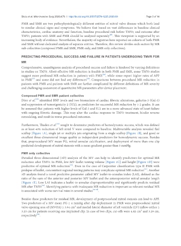
Furthermore, Thaden et al. sought to determine predictors of hemodynamic success, which was defined
as at least 40% reduction of left atrial V wave compared to baseline. Multivariable analysis revealed flail
scallop [Figure 1A], single jet or multiple jets originating from a single scallop [Figure 1B], and good or
excellent three-dimensional image quality as independent predictors for hemodynamic success. Besides
that, preprocedural MV mean PG, mitral annular calcification, and deployment of more than one clip
predicted development of mitral stenosis with a mean gradient greater than 5 mmHg.
PMR only collective
Detailed three-dimensional (3D) analysis of the MV can help to identify predictors for optimal MR
reduction after TMVr. In PMR, low MV leaflet tenting volume [Figure 1C] and height [Figure 1D] were
[31]
predictive of optimal MR reduction . Even in the case of Carpentier classification type II PMR with
[31]
prolapse of leaflet, concomitant regional tenting patterns may complicate optimal MR reduction . Another
3D analysis found a novel predictive parameter called MV leaflet-to-annulus index (LAI), defined as the
ratio of the sum of the anterior and posterior MV leaflet and the anteroposterior mitral annular length
[Figure 1E]. Low LAI indicates a leaflet-to-annulus disproportionality and significantly predicts residual
[32]
MR after TMVr . Identifying patients with inadequate MR reduction is important as relevant residual MR
is associated with worse survival rates in several studies [33-37] .
Besides these predictors for residual MR, development of postprocedural mitral stenosis can lead to APF.
Two predictors of a MV mean PG ≤ 4 mmHg after clip deployment in PMR were preprocedural mitral
valve opening area of (MVOA) ≥ 3.94 cm² and medial-lateral diameter of left ventricle (LV) inflow orifice ≥
3.23 cm for patients receiving one implanted clip. In case of two clips, cut-offs were 4.82 cm² and 3.29 cm,
[38]
respectively .