
Page 741 - Read Online
P. 741
Page 10 of 24 Palacios Mini-invasive Surg 2020;4:73 I http://dx.doi.org/10.20517/2574-1225.2020.72
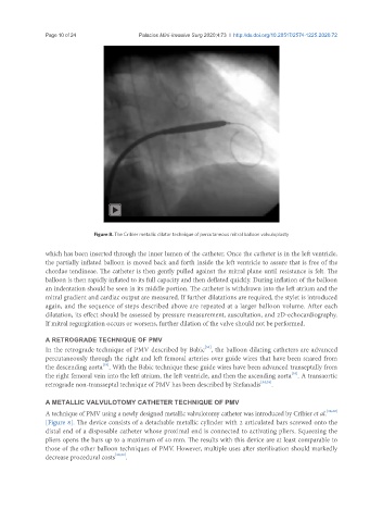
Figure 8. The Cribier metallic dilator technique of percutaneous mitral balloon valvuloplasty
which has been inserted through the inner lumen of the catheter. Once the catheter is in the left ventricle,
the partially inflated balloon is moved back and forth inside the left ventricle to assure that is free of the
chordae tendineae. The catheter is then gently pulled against the mitral plane until resistance is felt. The
balloon is then rapidly inflated to its full capacity and then deflated quickly. During inflation of the balloon
an indentation should be seen in its middle portion. The catheter is withdrawn into the left atrium and the
mitral gradient and cardiac output are measured. If further dilatations are required, the stylet is introduced
again, and the sequence of steps described above are repeated at a larger balloon volume. After each
dilatation, its effect should be assessed by pressure measurement, auscultation, and 2D-echocardiography.
If mitral regurgitation occurs or worsens, further dilation of the valve should not be performed.
A RETROGRADE TECHNIQUE OF PMV
[33]
In the retrograde technique of PMV described by Babic , the balloon dilating catheters are advanced
percutaneously through the right and left femoral arteries over guide wires that have been snared from
[33]
the descending aorta . With the Babic technique these guide wires have been advanced transeptally from
[33]
the right femoral vein into the left atrium, the left ventricle, and then the ascending aorta . A transaortic
retrograde non-transseptal technique of PMV has been described by Stefanadis [34,35] .
A METALLIC VALVULOTOMY CATHETER TECHNIQUE OF PMV
A technique of PMV using a newly designed metallic valvulotomy catheter was introduced by Cribier et al. [38,39]
[Figure 8]. The device consists of a detachable metallic cylinder with 2 articulated bars screwed onto the
distal end of a disposable catheter whose proximal end is connected to activating pliers. Squeezing the
pliers opens the bars up to a maximum of 40 mm. The results with this device are at least comparable to
those of the other balloon techniques of PMV. However, multiple uses after sterilization should markedly
decrease procedural costs [38,39] .