
Page 743 - Read Online
P. 743
Page 12 of 24 Palacios Mini-invasive Surg 2020;4:73 I http://dx.doi.org/10.20517/2574-1225.2020.72
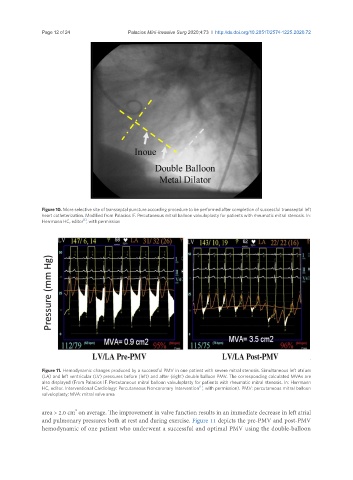
Figure 10. More selective site of transseptal puncture according procedure to be performed after completion of successful transseptal left
heart catheterization. Modified from Palacios IF. Percutaneous mitral balloon valvuloplasty for patients with rheumatic mitral stenosis. In:
[1]
Herrmann HC, editor , with permission
Figure 11. Hemodynamic changes produced by a successful PMV in one patient with severe mitral stenosis. Simultaneous left atrium
(LA) and left ventricular (LV) pressures before (left) and after (right) double balloon PMV. The corresponding calculated MVAs are
also displayed (From Palacios IF. Percutaneous mitral balloon valvuloplasty for patients with rheumatic mitral stenosis. In: Herrmann
[1]
HC, editor. Interventional Cardiology: Percutaneous Noncoronary Intervention , with permission). PMV: percutaneous mitral balloon
valvuloplasty; MVA: mitral valve area
2
area > 2.0 cm on average. The improvement in valve function results in an immediate decrease in left atrial
and pulmonary pressures both at rest and during exercise. Figure 11 depicts the pre-PMV and post-PMV
hemodynamic of one patient who underwent a successful and optimal PMV using the double-balloon