
Page 742 - Read Online
P. 742
Palacios Mini-invasive Surg 2020;4:73 I http://dx.doi.org/10.20517/2574-1225.2020.72 Page 11 of 24
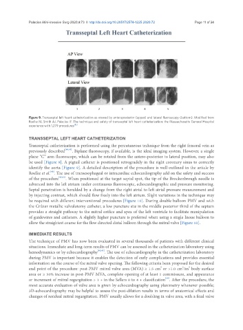
Figure 9. Transseptal left heart catheterization as viewed by anteroposterior (upper) and lateral fluoroscopy (bottom). Modified from
Roelke M, Smith AJ, Palacios IF. The technique and safety of transseptal left heart catheterization: the Massachusetts General Hospital
[51]
experience with 1,279 procedures
TRANSSEPTAL LEFT HEART CATHETERIZATION
Transseptal catheterization is performed using the percutaneous technique from the right femoral vein as
previously described [40,41] . Biplane fluoroscopy, if available, is the ideal imaging system. However, a single
plane “C” arm fluoroscope, which can be rotated from the antero-posterior to lateral position, may also
be used [Figure 9]. A pigtail catheter is positioned retrogradely in the right coronary sinus to correctly
identify the aorta [Figure 9]. A detailed description of the procedure is well outlined in the article by
[40]
Roelke et al. . The use of transesophageal or intracardiac echocardiography add on the safety and success
of the procedure [40,41] . When positioned at the target septal spot, the tip of the Brockenbrough needle is
advanced into the left atrium under continuous fluoroscopic, echocardiographic and pressure monitoring.
Septal penetration is heralded by a change from the right atrial to left atrial pressure measurement and
by injecting contrast, which should flow freely into the left atrium. Slight variations in the technique may
be required with different interventional procedures [Figure 10]. During double balloon PMV and with
the Cribier metallic valvulotomy catheter, a low puncture site in the middle posterior third of the septum
provides a straight pathway to the mitral orifice and apex of the left ventricle to facilitate manipulation
of guidewires and catheters. A slightly higher puncture is preferred when using a single Inoue balloon to
allow the straightest course for the flow directed distal balloon through the mitral valve [Figure 10].
IMMEDIATE RESULTS
The technique of PMV has now been evaluated in several thousands of patients with different clinical
situations. Immediate and long-term results of PMV can be assessed in the catheterization laboratory using
hemodynamics or by echocardiography [42,43] . The use of echocardiography in the catheterization laboratory
during PMV is important because it enables the detection of early complications and provides essential
information on the course of the mitral valve opening. The following criteria been proposed for the desired
2
2
2
end point of the procedure: post-PMV mitral valve area (MVA) ≥ 1.5 cm or >1.0 cm /m body surface
area or ≥ 50% increase in post-PMV MVA, complete opening of at least 1 commissure, and appearance
[39]
or increment of mitral regurgitation > 1 + in the Sellers 0 to 4 + classification . After the procedure, the
most accurate evaluation of valve area is given by echocardiography using planimetry whenever possible;
3D-echocardiography may be helpful to assess the post-dilation results in terms of anatomical effects and
changes of residual mitral regurgitation. PMV usually allows for a doubling in valve area, with a final valve