
Page 704 - Read Online
P. 704
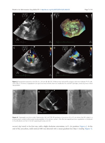
Rodinò et al. Mini-invasive Surg 2020;4:70  I  http://dx.doi.org/10.20517/2574-1225.2020.55                                    Page 7 of 10

                A                                             B


                C                                             D


               Figure 3. Preoperative evaluation with 2D (A, C, D) and 3D (B) TEE. White arrows indicate P2 prolapse; red arrow indicates P1-P2 cleft.
               Color TEE (C, D) shows 2 regurgitation jets (green arrows) with the main one localized at the level of P2 prolapse, producing severe mitral
               regurgitation


                A                           B                                  C


               Figure 4. Transseptal puncture under fluoroscopic (A) and TEE (B) guidance. A puncture 4 to 4.5 cm above the MV plane in a
               posterosuperior position allows good maneuverability of the delivery system. After the transseptal puncture, a guidewire is introduced
               in the left atrium and then in the left superior pulmonary vein (C)

               second clip lateral to the first one, with a slight clockwise orientation, in P1-A2 position [Figure 6]. At the
               end of the procedure, mild residual MR was detected with a mean gradient less than 5 mmHg [Figure 7].