
Page 700 - Read Online
P. 700
Rodinò et al. Mini-invasive Surg 2020;4:70 I http://dx.doi.org/10.20517/2574-1225.2020.55 Page 3 of 10
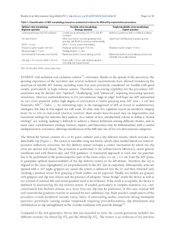
Table 1. Classification of MV morphology based on anatomical criteria for MitraClip implantation procedure
Optimal valve morphology Possible valve morphology Tough/unsuitable valve morphology
Beginner operator Average operator Expert operator
Central pathology (A2/P2 scallops) Commissural pathology (A1/P1 or A3/P3 Barlow’s syndrome, flail in multiple scallops
scallops)
No calcification Mild calcification outside grasping zone, Significant calcification of grasping zone
annulus calcification, previous annuloplasty
MVA > 4 cmq MVA > 3 cmq, preserved mobility -
Posterior leaflet length ≥ 10 mm Posterior leaflet length 7-10 mm Posterior leaflet length < 7 mm
Tenting height < 11 mm Tenting height > 11 mm -
Normal leaflet thickness and mobility Restricted leaflet motion during systole Restricted leaflet motion during systole and
diastole, rheumatic disease
Flail gap < 10 mm and width < 15 mm Flail width > 15 mm with dilated annulus -
(multiple clip implantation)
MV: mitral valve; MVA: mitral valve area
[13]
EVEREST trial inclusion and exclusion criteria ; nowadays, thanks to the spread of the procedure, the
growing experience of the operators and several technical improvements have allowed broadening the
spectrum of suitable MV lesions, including some that were previously considered not feasible with good
results, particularly in high-volume centers. Therefore, concerning eligibility for the procedure, MV
anatomies may be divided into “optimal”, “challenging” and “advanced”, requiring increasing operator
experience. Absolute contraindications to the percutaneous “edge-to-edge” technique are still represented
2
by very short posterior leaflet, high degree of calcification in leaflet grasping area, MV area < 3 cm and
[14]
rheumatic MR [Table 1]. An interesting topic is the management of MR of mixed or undetermined
etiologies, but data in this regard are still scant. To date, only few registries report an incidence ranging
from 3% to 10% in real-world practice; however, their results have focused only on degenerative and
functional etiology for outcome data analysis. As a matter of fact, standardized criteria to define a “mixed
etiology” are lacking, making it difficult to achieve a shared definition among different studies, and in
most cases a predominant etiology between organic and functional may be individualized with a careful
multiparametric evaluation, allowing classification of the MR into one of the two dichotomous categories.
The MitraClip System consists of a 24 Fr guide catheter and a clip delivery system, which includes one
detachable clip [Figure 1]. The system is steerable using two knobs, which allow medial-lateral and anterior-
posterior deflection; moreover, the clip delivery system includes a control mechanism by which the clip
arms are opened and closed. The procedure is performed in the catheterization laboratory, under general
anesthesia and with fluoroscopic and TEE guidance. A transseptal approach is used, and the puncture
has to be performed in the posterosuperior part of the fossa ovalis, 4.5 cm ± 0.5 cm from the MV plane,
to guarantee optimal maneuverability of the clip delivery system in the left atrium. Therefore, the clip is
aligned on the main regurgitation jet, perpendicularly to the MV line of coaptation. Subsequently, arms are
opened with a 180° angle, grippers are raised, the system is advanced into the LV and then retracted until
reaching a position where firm grasping of both leaflets can be expected. Finally, the leaflets are grasped
with grippers and clip arm closure and the presence of adequate “tissue-bridge” inside the device as well as
the amount of residual MR and mitral gradient need to be evaluated. If the result is acceptable, the device is
deployed by maneuvering the clip delivery system. If needed, particularly in complex anatomies (i.e., cleft,
commissural flail, Barlow’s disease, etc.), more than one clip may be positioned. In this case, residual MR
and transvalvular gradients must be re-assessed for each additional clip. Main possible complications of the
procedure include peripheral vascular injury, injury of surrounding cardiac structures during transseptal
puncture, potentially causing cardiac tamponade requiring pericardiocentesis, clip detachment and
[15]
embolization or clip entanglement in the chordae tendineae with possible damage .
Compared to the first-generation device that was launched in 2008, the current generation includes two
different versions: the MitraClip NT and the MitraClip XT . The former is an evolution of the previous
R
R