
Page 221 - Read Online
P. 221
Mok et al. Mini-invasive Surg 2020;4:26 I http://dx.doi.org/10.20517/2574-1225.2020.04 Page 5 of 6
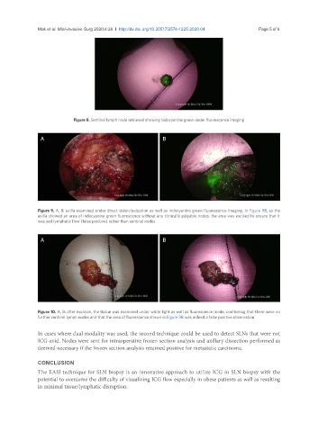
Figure 8. Sentinel lymph node retrieved showing indocyanine green under fluorescence imaging
A B
Figure 9. A, B: axilla examined under direct vision/palpation as well as indocyanine green fluorescence imaging. In Figure 9B, as the
axilla showed an area of indocyanine green fluorescence without any clinically palpable nodes, the area was excised to ensure that it
was just lymphatic flow (false positive) rather than sentinel nodes
A B
Figure 10. A, B: after excision, the tissue was examined under white light as well as fluorescence mode, confirming that there were no
further sentinel lymph nodes and that the area of fluorescence shown in Figure 9B was indeed a false positive observation
In cases where dual modality was used, the second technique could be used to detect SLNs that were not
ICG-avid. Nodes were sent for intraoperative frozen section analysis and axillary dissection performed as
deemed necessary if the frozen section analysis returned positive for metastatic carcinoma.
CONCLUSION
The EASI technique for SLN biopsy is an innovative approach to utilize ICG in SLN biopsy with the
potential to overcome the difficulty of visualizing ICG flow especially in obese patients as well as resulting
in minimal tissue/lymphatic disruption.