
Page 218 - Read Online
P. 218
Page 2 of 6 Mok et al. Mini-invasive Surg 2020;4:26 I http://dx.doi.org/10.20517/2574-1225.2020.04
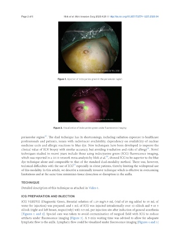
Figure 1. Injection of indocyanine green in the periareolar region
Figure 2. Visualization of indocyanine green under fluorescence imaging
[1]
periareolar region . The dual technique has its shortcomings, including radiation exposure to healthcare
professionals and patients, issues with radiotracer availability, dependency on availability of nuclear
medicine units and allergic reactions to blue dye. New techniques have been developed to improve the
[2]
clinical value of SLN biopsy with similar accuracy, but avoiding irradiation and risks of allergy . Novel
techniques studied in recent years include those using indocyanine green (ICG) fluorescence imaging,
[3]
which was reported in a 2019 network meta-analysis by Mok et al. , showed ICG to be superior to the blue
dye technique alone and comparable to that of the standard dual-modality method. There was, however,
[4]
technical difficulties with the use of ICG especially in obese patients, thereby limiting the widespread use
of this modality. In this article, we describe a minimally invasive technique which is effective in overcoming
limitations and at the same time minimizes tissue dissection or disruption in the axilla.
TECHNIQUE
Detailed description of this technique as attached in Video 1.
ICG PREPARATION AND INJECTION
ICG VERDYE (Diagnostic Green, Bavaria) solution of 1.25 mg/0.5 mL (vial of 25 mg added to 10 mL of
water for injection) was prepared, and 1 mL of ICG was injected intradermally over 12 o’clock and 9 or 3
o’clock (right and left breast, respectively) with 0.5 mL per injection site after induction of general anesthesia
[Figures 1 and 2]. Special care was taken to avoid contamination of surgical field with ICG to reduce
artifacts under fluorescence imaging [Figure 3]. A 5-min waiting time was advised to allow for adequate
lymphatic flow to the axilla. Lymphatic flow could be visualized under fluorescence imaging [Figures 4 and 5]