
Page 72 - Read Online
P. 72
Shimada et al. Mini-invasive Surg 2019;3:7 I http://dx.doi.org/10.20517/2574-1225.2018.78 Page 5 of 11
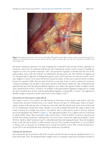
Figure 1. We present the boundary of No.6 lymph node station. The operator should identify anterior superior pancreaticoduodenal vein
(ASPDV) and elevates No.6 lymphoid tissues ventrally and softly. ARCV: accessory right colic vein; GCT: gastrocolic trunk; RGEV: right
gastroepiploic vein
the greater omentum; moreover, the wide triangulation is formed by the traction of fatty appendices of
transverse colon with the assistant’s left forceps. The triangulated counter traction makes it possible for
surgeons to resect the greater omentum. Then, the splenocolic ligament is divided and the root of left
gastroepiploic artery and vein (LGEAV) are identified in the pancreatic tail. After LGEAV are ligated and
cut, the gastrosplenic ligament including short gastric artery (SGA) and vein are dissected toward cranial
side. Surgeon is careful not to injure the branch around the spleen. In the area of superior border of spleen,
because the operative field often becomes limited by excess fatty tissue, in such occasions the precursor
cutting of esophagus may be able to show good operative field toward caudal side later. Alternatively the
approach that goes into the left gastrophrenic ligament can confirm the superior border of spleen and the
most cranial branch of SGA. Therefore, the mobility of the gastrosplenic ligament is improved by cutting
the most cranial branch of SGA, and the gastrosplenic ligament is spread like a “screen”. This approach is
[21]
flexible enough to respond to variety of pancreatic tail or splenic hilum .
Dissection of infra-pyloric nodes (No.6)
The surgeon moves to the patient’s left side during the dissection of No.6 lymph node station. After the
omentectomy has been finished close to the hepatic flexure, the layer of “embryologic failure of fusion”
which consists of the anterior lobe of transverse mesocolon and the anterior pancreatic fascia is dissected
for the mobilization of transverse colon. Thereby, we are able to finally determine the range to dissect and
ready to dissect No.6 lymph node station. The inferior limit of infra-pyloric nodes is anterior superior
pancreaticoduodenal vein (ASPDV) [Figure 1]. The right gastroepiploic vein is clipped and cut at the point
in which ASPDV flows. Then, the lymph nodes and fat tissue in front of ASPDV is elevated and dissected
toward descending duodenum. Subsequently, the neural layer around the right gastroepiploic artery
(RGEA), which is called “outermost layer”, is identified and dissected keeping the layer. Then, the lymphoid
tissue on the left side of RGEA is softly elevated ventrally. Nerve bundle around RGEA is cut and RGEA
is clipped and cut. Because the lymphoid tissue around infra-pyloric artery (IPA) remain formed into a
screen, IPA is cut and lymphoid tissue attaching to duodenum is peeled up.
Cutting the duodenum
The assistant lift up the posterior wall of the stomach, and the branches of superior duodenal artery is cut
from dorsal side. Then, the duodenal bulb is stapled and cut. In patients suspected of duodenal invasion of