
Page 314 - Read Online
P. 314
Mok et al. Mini-invasive Surg 2019;3:40 I http://dx.doi.org/10.20517/2574-1225.2019.39 Page 5 of 9
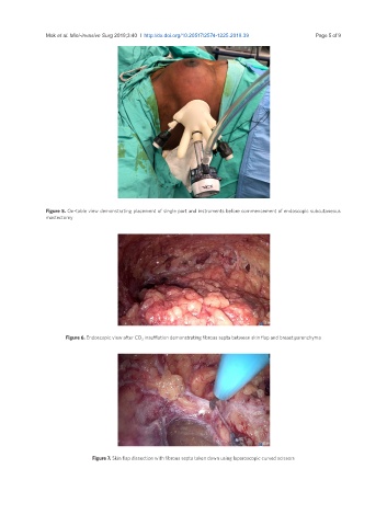
Figure 5. On-table view demonstrating placement of single port and instruments before commencement of endoscopic subcutaneous
mastectomy
Figure 6. Endoscopic view after CO 2 insufflation demonstrating fibrous septa between skin flap and breast parenchyma
Figure 7. Skin flap dissection with fibrous septa taken down using laparoscopic curved scissors